TETHERED SPINAL CORD : PART 3 SPINAL DYSRAPHISM
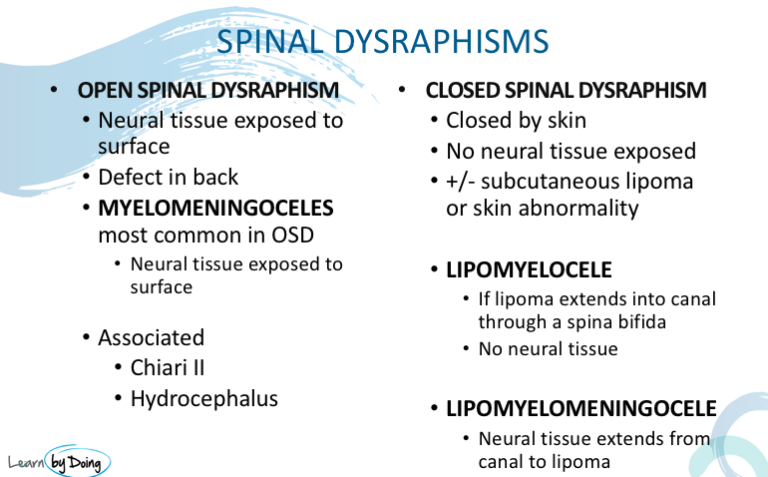
- There are many Spinal Dysraphisms which are malformations of the cord and spine. It gets fairly confusing so start by dividing them into eithr
- Open ( neural tissue exposed to view through skin defect) and
- Closed ( malformation covered by skin and not open to view) dysraphisms.
- If its Open, the most most common type of Open Spinal Dysraphism is a Myelomeningocele
- If its a Closed Dysraphism, these can be divided into those with a subcutaneous mass and without.
- With Subcutaneous Mass ( Lipoma): Most common are Lipomyelocele and Lipomyelomeningocele. Less common are meningoceles and myelocystoceles.
- Without Subcutaneous Mass:
- Simple: Filum fat/ lipoma, Intradural lipomas, Persistent ventriculus terminalis Dermal sinuses
- Complex: Diastematomyelia, Sacral agenesis
Image Above: Diagram demonstrates a Myelomeningocele. Neural placode ( * and red) protruding through skin ( brown) together with CSF and arachnoid ( blue).
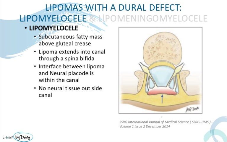
 Image Above: Diagram demonstrates a Lipomyelocele. Skin ( brown) is closed . Subcutaneous lipomatous mass ( yellow) is attached to the neural placode ( red) within the canal.
 Image Above: Diagram demonstrates a Lipomeningomyelocele. Skin ( brown) is closed . Subcutaneous lipomatous mass ( yellow) is attached to the neural placode ( red) outside the canal.


