Tethered Cord Syndrome
Tethered Cord Syndrome is where the
- Conus is low lying and the
- Filum terminale is tethered.
This can be caused by a number of spinal malformations and can present at any age.
The pathophysiology behind the clinical symptoms is that the tight, tethered filum pulls and stretches the conus resulting in ischaemia to the conus.
Before we look at the various causes of tethered cord syndrome lets look at the normal Conus and Filum.
Normal Conus
|
- The cord grows at a slower rate than the vertebral column.
- At birth the normal conus lies at the L2/3 level and the thecal sac is at the S2 level.
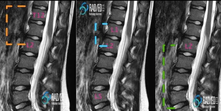
- By 2/12 the conus usually lies around the L1/2 level ( blue) .
- Conus at or above mid L2 is normal at any age ( orange).
- Conus below mid-L2 abnormal ( green) : This is the statement from the International Society of Paediatric Neurosurgeons. ” Any conus medullaris lying caudal to the mid-body of L2 is considered abnormally low (95% confidence limits) and therefore potentially tethered.”

Image Above: Range of locations for the conus. See text above image for explanation.
|
Normal Filum
|
- The filum terminale is a continuation of the cord and arises from the pial layer.
- It attaches proximally to the conus ( intra dural) and distally to the first coccygeal segment ( extra dural)
- It is predominantly fibrous.
- Normal size is approximately 2mm in width proximally but becomes thinner distally.
- Usually not seen on MRI.

Image Above: Normal Filum Terminale ( orange arrow). Low signal on T2 in midline. |


