MRI of Postero Medial Ankle Impingement (POMI)
Pathology: What causes the impingement
|
|
Postero Medial Ankle Impingement is usually secondary to contusion/ tearing of the posterior fibres of the deep deltoid ligament ( the Posterior Tibio Talar Ligament) and the postero medial capsule.
This results in hypertrophic scarring and fibrosis which extends into the postero medial gutter between the FDL and FDH tendons ( see anatomy below) resulting in impingement .
|
Anatomy: How to Find the Postero Medial Recess
|
|
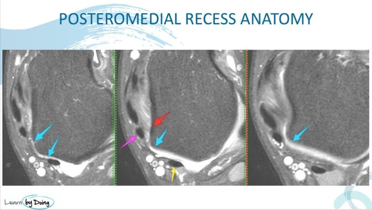
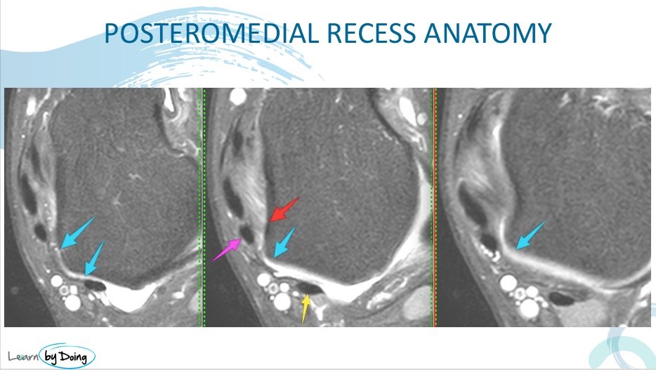
 Image Above: Demonstrates where to find the posteromedial gutter or recess ( Blue arrows). The key if to find the space between the FDL and FHL where the recess lies. Image Above: Demonstrates where to find the posteromedial gutter or recess ( Blue arrows). The key if to find the space between the FDL and FHL where the recess lies.
- ANTERIOR MARGIN:
- Medial Malleolus and Posterior fibres Deep Deltoid ( Red arrow )
- POSTERIOR: Capsule
- Recess lies between FDL ( pink arrow) and FHL ( yellow arrow)
|
| What Do You Look For on MRI? |
LOOK IN POSTEROMEDIAL RECESS FOR
- Scar, synovitis or thickening in recess between FDL and FHL
- Posterior capsular thickening at level of recess
- Torn Posterior deep deltoid
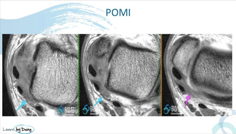
 Image Above: Thickening secondary to granulation tissue/ scar in the posteromedial recess ( pink and blue arrows). Key is to find the space between FDL and FHL to identify the region of the postero medial recess. The deep deltoid fibres ( not arrowed) anterior to the recess are ill defined and torn. Image Above: Thickening secondary to granulation tissue/ scar in the posteromedial recess ( pink and blue arrows). Key is to find the space between FDL and FHL to identify the region of the postero medial recess. The deep deltoid fibres ( not arrowed) anterior to the recess are ill defined and torn.
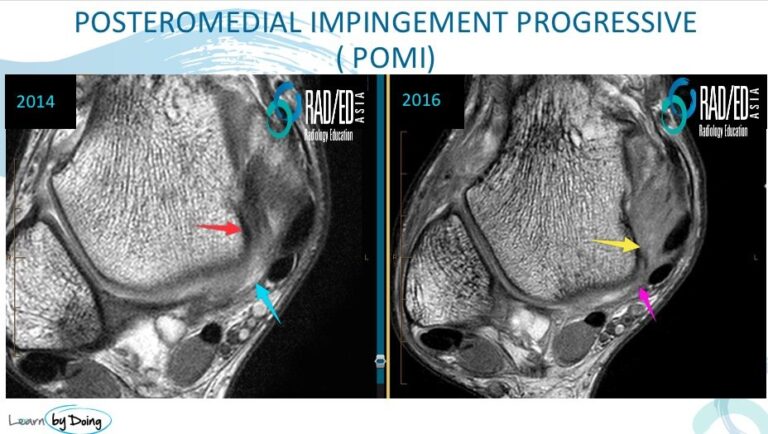
 Image Above: First image demonstrates normal posteromedial recess ( blue arrow) and normal posterior deep deltoid ( red arrow). 2nd Image two years post trauma very faint filling of the postero medial recess with granulation tissue/ scar ( pink arrow) and ill definition of the posterior fibres of the deep deltoid ( yellow arrow). Image Above: First image demonstrates normal posteromedial recess ( blue arrow) and normal posterior deep deltoid ( red arrow). 2nd Image two years post trauma very faint filling of the postero medial recess with granulation tissue/ scar ( pink arrow) and ill definition of the posterior fibres of the deep deltoid ( yellow arrow).
|


