MRI Findings in Posterior Ankle Impingement
Intro: In this post we are going to look at the MRI findings that can be seen with Posterior Impingement. If you havent read the post on the bony changes in the posterior ankle that can predispose to impingement please read that first here at this link
Its also important to recognise that that we can only report on the findings that suggest impingement and not say that the patient HAS impingement. This is something for the clinician to determine with the clinical and radiological findings.
| Need to Know: Pathology |
|
Look for the causes/ sequelae of impingement listed below. In addition look for thickening/ ill definition or scarring of the PTFL and the Intermalleolar ligament.

|
| MRI Findings in Impingement |
|

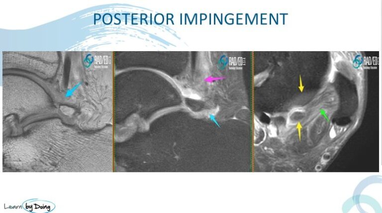
Image Above:Findings suggest posterior impingement. Posterior synovitis ( blue arrows), Soft tissue oedema ( pink arrow), Os trigonum with oedema ( yellow arrows) and ill definition and hyperintensity of the PTFL ( green arrow).
 Image Above: Thick hyperintense PTFL ( Blue arrows) and unstable os trigonum with fluid in syndesmosis ( yellow arrow) and bone oedema ( pink arrows). Image Above: Thick hyperintense PTFL ( Blue arrows) and unstable os trigonum with fluid in syndesmosis ( yellow arrow) and bone oedema ( pink arrows).
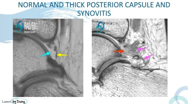
 Image Above: Normal posterior capsule ( yellow arrow) and normal posterior joint fluid ( blue arrow). Compare with capsular thickening ( pink arrows) and synovitis ( red arrow) where the joint fluid signal is reduced. Image Above: Normal posterior capsule ( yellow arrow) and normal posterior joint fluid ( blue arrow). Compare with capsular thickening ( pink arrows) and synovitis ( red arrow) where the joint fluid signal is reduced.
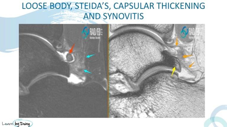
 Image Above: Findings of a loose body ( red arrow), Steida’s Process ( yellow arrow), capsular thickening and synovitis ( orange arrows) and peri capsular oedema ( blue arrows) suggest posterior impingement. Image Above: Findings of a loose body ( red arrow), Steida’s Process ( yellow arrow), capsular thickening and synovitis ( orange arrows) and peri capsular oedema ( blue arrows) suggest posterior impingement.
|


