MRI Wrist TFCC Anatomy
The MRI anatomy of the TFCC is important to know before we start talking about TFCC abnormalities. Today’s post is on the normal anatomy of the TFCC which is composed of a number of structures.
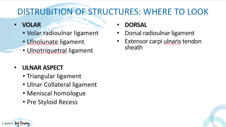
Image Above: Triangular fibrocartilage ( blue arrow) also called articular disc. Ulnar side thicker than radial. Radial side attaches to radial articular cartilage. Black on PD and PDFS. Is biconcave in appearance being thinner centrally. In diagram ( credit see below) TFC= Triangular Fibro Cartilage, UL= Ulno Lunate Ligament, UT = Ulno Triquetral Ligament, *= Pre Styloid Recess, MH= Meniscal Homologue, CL= Ulnar Collateral Ligament,
Image Above: Diagram is looking from above. TFC Fuses with Volar and Dorsal RadioUlnar Ligaments on either side.
Image Above: Triangular ligaments are the ulnar attachment of the TFC to the ulnar styloid ( red arrow) and the fovea ( ligament yellow arrow, fovea pink arrow). In between the foveal and styloid attachments is the Ligamentum Subcruentem.
 Image Above: Pre styloid recess ( red arrow) Meniscal Homologue ( blue arrow) ill defined thickening arises from ulnar styloid and merges with Triquetrum. ECU ( yellow arrow).
 Image Above: Ulnolunate ( blue arrow sagittal scan) and Ulnotriquetral ( pink arrow coronal and sagittal). Difficult to see on coronal scans as they run in plane. Can be easier on sagittal. Combination of Ulnolunate, Ulnotriquetral and UlnoCapitate ligaments called Ulnocarpal ligamentous complex. Cant distinguish one from another as they are fused.
Ulnar Collateral Ligament is mentioned in texts but is mostly not seen on MRI. Arises from tip of ulnar styloid.
All diagrams from Zhan HL et al. High‐resolution 3T Magnetic Resonance Imaging of the Triangular Fibrocartilage Complex in Chinese Wrists: Correlation with Cross‐sectional Anatomy. Chin Med J 2017;130:817‐22.


