msk mri radiology conference

MRI Wrist Tendinosis and Tenosynovitis

Tendinosis and Tenosynovitis on MRI of the wrist have a common appearance with varying amounts of enhancement, fluid and tendon hyperintensity. The appearance is common to all tendons in the wrist. 2 minute read on the various appearances on MRI.

mri wrist tendinosis tenosynovitis radiology education asia

mri wrist tendinosis tenosynovitis enhancement radiology education asia

Image Above: Tenosynovitis and tendinosis. Thick walled ( blue arrow) with enhancement ( yellow arrow) and fluid ( red arrow) in tendon sheath.

mri wrist tendinosis tenosynovitis fluid enhancement radiology education asia

Image Above: Tenosynovitis and tendinosis. Thick low T2FS signal wall ( blue arrow) with enhancement ( yellow arrow) and fluid ( red arrow) in tendon sheath.

mri wrist tendinosis tenosynovitis fluid tendon sheath radiology education asia


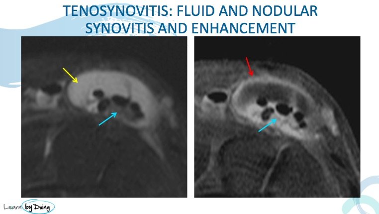
Image Above: Tenosynovitis and tendinosis. Nodular synovial enhancement which is low T2 signal within the tendon sheath ( blue arrow), enhancing tendon sheath ( red arrow) and fluid ( yellow arrow) in tendon sheath.


mri wrist tendinosis tenosynovitis bone oedema fluid enhancement radiology education asia

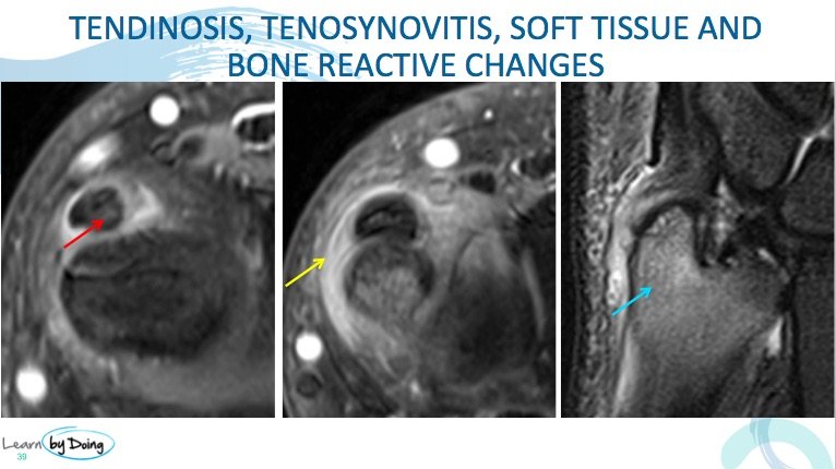
Image Above: Tenosynovitis and tendinosis. Soft tissue thickening and enhancement ( yellow arrow) and increased tendon signal ( red arrow). Reactive bone marrow oedema ( blue arrow) in ulnar.

Leave a Comment

Radiology Education

Join our Newsletter

Stay tuned on new
Mini-Fellowships launches and learnings

Mini-Fellowships

Better Than Conferences

We help radiologists learn by doing and report with confidence

Scroll to Top

Log in to your Mini-Fellowship or create an account

Mini-Fellowships

Better Than Conferences

We help radiologists
learn by doing and
report with confidence

Join our Newsletter

Stay tuned on new
Mini-Fellowships launches and learnings

Mini-Fellowships

Better Than Conferences

Join our WhatsApp Community

We help radiologists learn
by doing and report with confidence