MRI Wrist Scapho Lunate Disassociation
Scapho lunate disassociation is the separation of the the lunate and scaphoid due to a significant tear of the scapholunate ligament. This leads to instability and premature OA. We saw in the workshop and in previous posts what SLL tears look like on MRI. This post is a brief overview on what SL disassociation looks like.
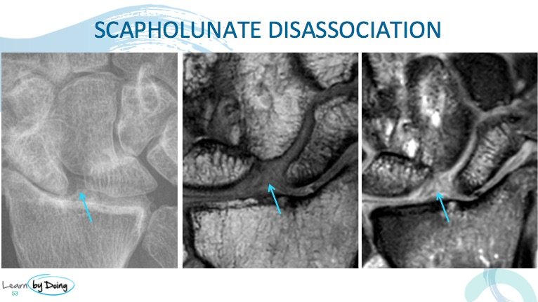
Image Above: Tear of the SLL leads to a widening of the scapholunate joint of greater than 3mm( blue arrow) best assessed on the coronal scans.
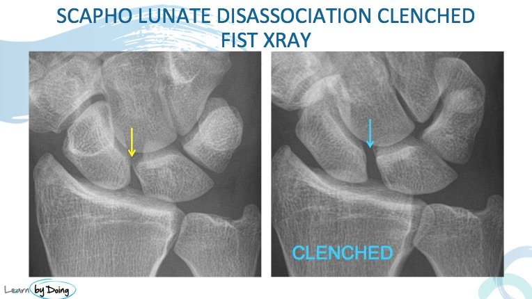
Image Above: Widening of the scapholunate joint may only be seen with the SL joint stressed as in a clenched fist view ( blue arrow).
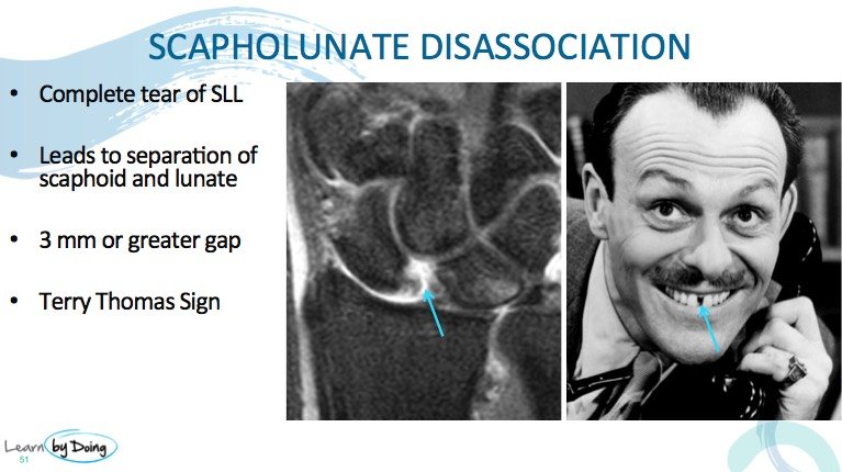
 Image Above: Tear of the SLL widening of the scapholunate joint ( blue arrow) xray and MRI.


