MRI Wrist Pre Styloid Recess Synovitis
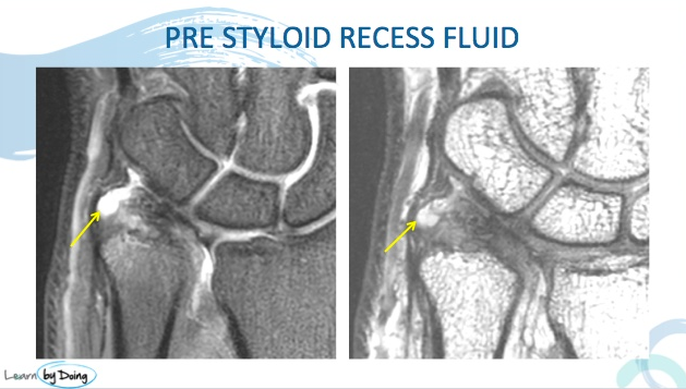
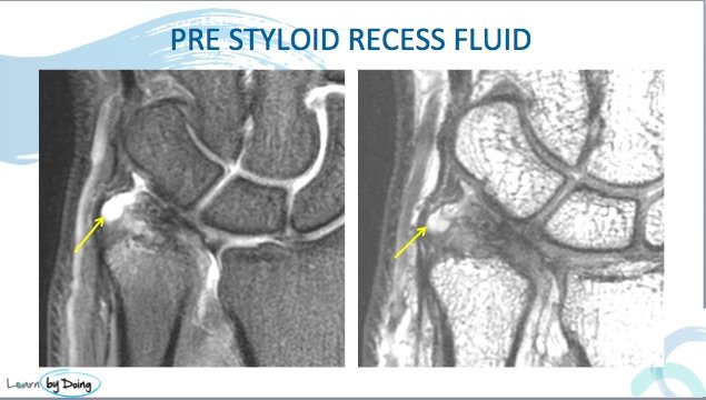
The Pre Styloid Recess lies adjacent to the ulnar styloid and TFC. The clinical significance of it is that it can become inflamed and be a cause of pain. This post looks at the MRI appearance of Synovitis and Fluid in the Pre Styloid Recess.
 Image Above: Location of pre styloid recess ( red arrow). Meniscal homologue ( blue arrow). ECU tendon ( yellow arrow)Diagram credit below.
 Image Above: Pre Styloid Recess fluid (yellow arrow), synovitis ( blue arrow) and styloid process ( red arrow).
 Image Above: Styloid process (blue arrow) with synovial thickening and fluid volar to it ( yellow arrow).
 Image Above: Fluid in the pre styloid recess ( yellow arrow).
Diagram from Zhan HL et al. High‐resolution 3T Magnetic Resonance Imaging of the Triangular Fibrocartilage Complex in Chinese Wrists: Correlation with Cross‐sectional Anatomy. Chin Med J 2017;130:817‐22.


