Ulnar Impaction or Abutment is when the head of the ulnar impinges on more distal structures resulting in degeneration, tears and pain. One of the causes of Ulnar Abutment is Positive ulnar variance where the ulnar is too long relative to the radial articular surface at the wrist.
WHAT ARE THE CAUSES OF ULNAR ABUTMENT AND WHAT EFFECT DOES IT HAVE?

HOW TO DEFINE ULNAR VARIANCE
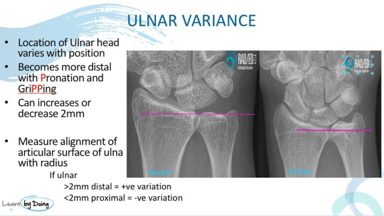
Draw a horizontal line across the radial articular surface adjacent to the DRUJ. Then measure the distance of the head of the ulnar from that line. If its more than 2mm its considered Ulnar Positive. If its less than 2mm its Ulnar Negative. In between is Ulnar Neutral.
You need to be aware that in a normal wrist, the position of the ulnar head relative to the radial articular surface can increase by upto 2mm with Pronation and Gripping. So you can have someone who on xray is Ulnar Neutral but has signs of Ulnar impaction. You can further assess this by doing the xray in pronation and with gripping to see if the ulnar becomes positive.
WHAT DOES IT LOOK LIKE ON MRI
Look for these things that suggest Impaction. Remember that the ulnar can be neutral in location and there can still be abutment as the ulnar moves with pronation and gripping ( our MRI is done at rest in a neutral position). So if you see other signs of Impingement but a neutral ulnar, it can still be Ulnar Impaction.
- Positive Ulnar Variance
- Oedema / sclerosis in the ulnar head/ lunate or less commonly the triquetrum.
- TFC: Thinning, delamination and tearing
- Chondral loss over the lunate , ulnar or triquetrum
- TFC: Scarring/ delamination TFC attachments to ulnar styloid or meniscal homologue. ( Less common)
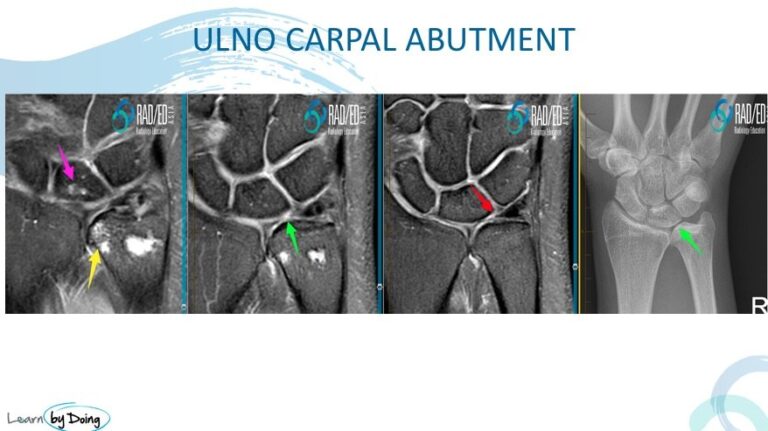
Image Above: Full range of findings suggesting ulnar impaction. Ulnar positive ( green arrow in xray). TFC tear ( green arrow in MRI), Cartilage loss ulnar aspect of lunate ( red arrow), Subcortical cystic degenerative changes in lunate ( purple arrow) Degenerative change sin DRUJ ( yellow arrow).
Image Above: Positive Ulnar Variation ( blue arrow). Oedema in lunate ( red arrow) with thinning of lunate cartilage ( pink arrow) and thinning of TFC ( orange arrow).

Image Above: Positive Ulnar Variation ( blue arrow). Oedema and cyst formation in ulnar head ( red arrows) with scarring of ulnar and meniscal homologue attachments of TFC ( yellow arrow).

Image Above: Neutral Ulnar Variation ( blue arrow). Oedema in lunate ( orange arrow) with thinning of TFC ( yellow arrow).


