MRI TENDONS: AN EASY PATTERN TO ASSESS
HOW DO TENDONS RESPOND TO INJURY
MRI TENDONS: How do tendons respond to injury?
MRI TENDONS : MSK MRI requires you need to have a good understanding of radiological anatomy, but one of the best things about MSK MRI is that the response to injury is similar if not the same in most joints. So often you can learn the response in one area and apply it to every other joint. So what’s the response to injury seen in MRI Tendons?
It’s pretty limited.
The options are to gather some fluid, become small, big, bright, really bright or just disappear.
Start with knowing the Normal MRI of Tendons.
MRI Normal tendon: Uniformly black on T1/T2/PD scans (Medial Ankle Tendons). A minor amount of fluid in the tendon sheath can be normal.
MRI Tenosynovitis: Fluid in the tendon sheath A minor amount of fluid in the tendon sheath such as in the normal example at the beginning, is alright. However an increased amount of fluid (blue arrow) indicates inflammatory changes of the tendon sheath. The tendon itself may be normal. In the example above, the Tibialis Posterior tendon (anterior most tendon) is moderately enlarged.
MRI Tendinosis: Increase in Tendon size and or Increase in Tendon Signal. Significantly increase in size of the Tibialis Posterior tendon (blue arrow). There is also mild increased intermediate signal in the tendon but the signal is not the same as fluid signal, indicating tendinosis rather than a tear. Note significant tenosynovitis.
MRI Partial Tear: Split Tendon. The tibialis posterior tendon (Orange arrow) is split (Pink arrow) in two (double blue arrows). Single tendon before and after the split. Mild increased signal before and after split indicates tendinosis.
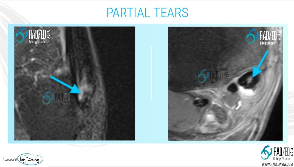
MRI Partial Tear: Fluid signal within the tendon (Blue arrow) indicates a tear of the tendon fibres. Once something is torn there cant be an empty space, something has to fill it, and it gets filled initially with fluid. Note that the signal intensity is much higher than tendinosis.
MRI Complete Tendon Tear : There is complete loss of tendon fibres and the area is filled with fluid. In the shoulder above the pink arrow indicates the tear filled with fluid. The orange arrow indicates tendinosis which is of a less bright signal. Complete rupture of the biceps tendon with an empty tendon sheath (Blue arrow).
MRI Peritendonitis : For tendons such as the Achilles or Gluteal tendons that don’t have a tendon sheath, inflammatory changes spread into the adjacent soft tissues (Blue arrow high signal in pre Achilles fat).
That’s it. Pretty simple and repetitive pattern which can be applied to any tendon.
VIEW VIDEO
https://youtu.be/jCP89RCRwU0
If your Browser is blocking the video, Please view it on our YouTube Channel HERE.
Learn more about MRI Imaging in our ONLINE or ONSITE
Guided MRI Mini-Fellowships.
More by clicking on the images below.


VIEW OUR LATEST POPULAR BLOG-POSTS: CLICK ON THE IMAGES BELOW
#radedasia
#radiology #radedasia #radiologist #radiologia #mri #mskmri #spinemri #msk #mskrad #mskradiology #imaging #frcr #sportsmed #radiologyresident #foamrad #ortho #ct #radiologystudent #trauma #radedasia #radiologycme #radiologyeducation #radiologycases #rheumatology #arthritis #painphysician #physiotherapy #spondyloarthropathies #chiropractic


