MRI Knee : Menisco Capsular Separation
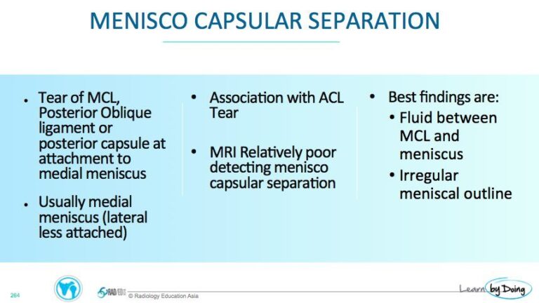
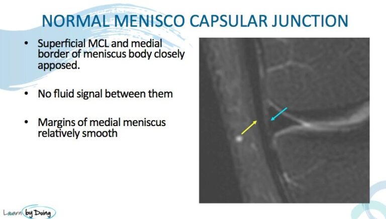
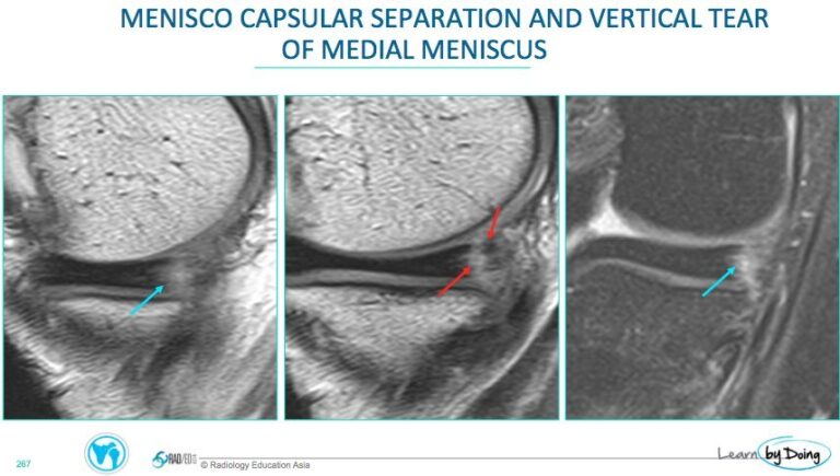
What do you assess when you are looking for menisco capsular separation on MRI? The superficial MCL/ posterior oblique ligament and the medial margin of the body of the medial meniscus are closely opposed and can be torn apart with trauma. When they separate, fluid can accumulate in the space between them and the meniscal margin can be ripped resulting in a serrated appearance (think of the appearance of skin peeled off from a band aid removal). The posterior horn of the medial meniscus adjacent to the separation also can be torn ( commonly a vertical tear).
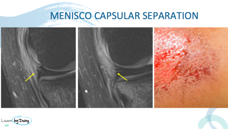
Image Above: Meniscocapsular separation very irregular margins of the body of the medial meniscus and high T2 signal adjacent to meniscus ( yellow arrow). Meniscal surface is like a band aid has been peeled off the skin ( last image).
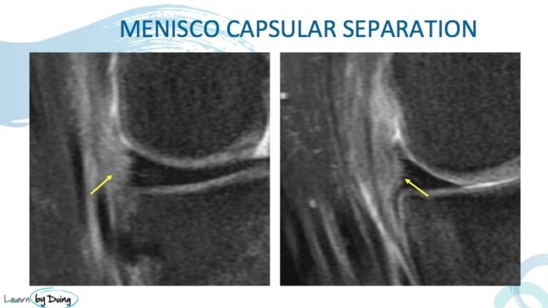
Image Above: Meniscocapsular separation very irregular margins of the body of the medial meniscus ( yellow arrow).
Image above: 1st and 2nd images sagittal, 3rd image coronal. Meniscocapsular separation ( blue arrows 1st and 3rd images). Peripheral vertical tearing of the adjacent posterior horn of the medial meniscus ( red arrows).

