MRI Knee Fat Pad Impingement : Quadriceps, Supra Patella and Hoffa’s Fat Pads
There are three fat pads around the knee and signs of impingement can be seen on MRI in all three. 2 minute review of the MRI changes of fat pad impingement.
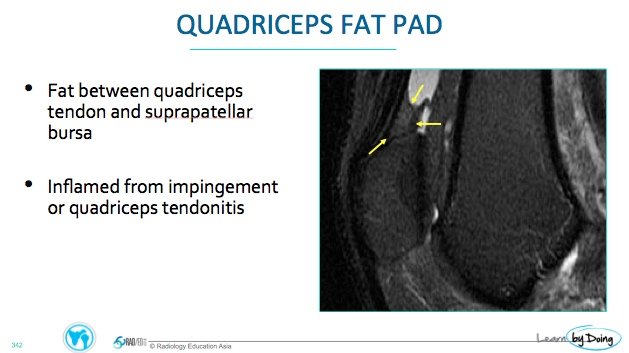
MRI APPEARANCE OF IMPINGEMENT: Increased T2/PDFS signal in the fat pad affected. It can be swollen and have convex margins particularly the quadriceps fat pad.
QUADRICEPS FAT PAD:
 Image above: PDFS Quadriceps fat pad lies immediately posterior to the quadriceps insertion. It should not have high signal on PDFS and its posterior margin is usually concave.
 Image Above: Quadriceps fat pad impingement with increased signal on PDFS ( yellow arrow) and convex posterior margin ( blue arrow).
SUPRAPATELLAR FAT PAD:
HOFFA’S FAT PAD:


