MRI ACL Mucoid Degeneration
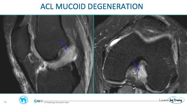
Mucoid Degeneration of the Anterior Cruciate Ligament (ACL ) can mimic a tear on MRI. Review of the findings of mucoid degeneration on MRI
 Image Above: ACL mucoid degeneration. Expansion of the ACL but ACL fibres are still seen in normal alignment.
Image Above: Complete tear ( left ) and mucoid degeneration ( right). In mucoid degeneration ACL fibres are still seen in normal alignment and continuity and cysts are present which would not be seen in a tear.
 Image Above: ACL mucoid degeneration. Expansion of the ACL but ACL fibres are still seen in normal alignment. Cyst ( yellow arrow) present adjacent to the tibial ACL insertion site and bone marrow oedema ( blue arrow) at the insertion..
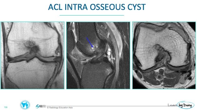
 Image Above: ACL mucoid degeneration. ACL not expanded and ACL fibres in normal alignment. Intraosseous cyst and oedema adjacent to the femoral attachement site of the ACL.


