MRI Hip Subchondral Insufficiency Fractures
Subchondral Insufficiency Fractures of the Hip on MRI have a specific appearance which is useful in differentiating them from AVN of the hip.
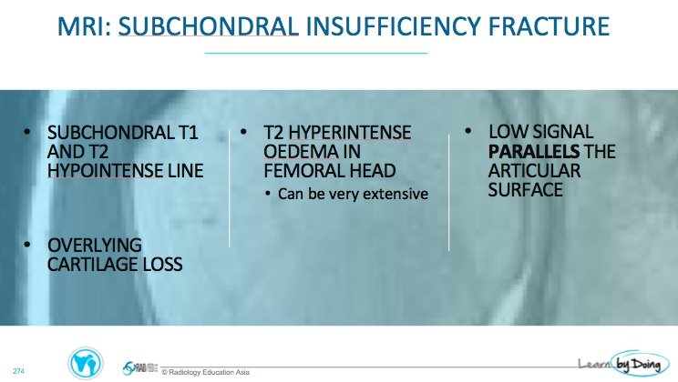
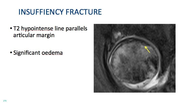
Like all fractures on MRI they are low signal on all sequences with surrounding bone marrow oedema. The important thing is that they PARALLEL the cortex and their inferior margin is concave. In time, they either heal and sclerose ( low signal on all sequences) or the cortex collapses. Here is what they look like
Image Above: Blue arrow is cortex. The Yellow Arrows indicate the subchondral insufficiency fracture. Note how it parallels the line of the cortex and its inferior margin is concave.
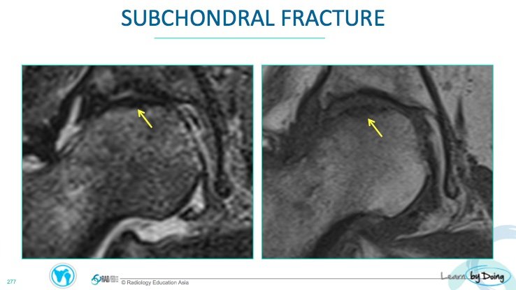
Image Above: Subchondral insufficiency fracture ( yellow arrow) on T2FS (left image) and T1 ( right image). The fracture line is very close to the cortex and sometimes what we see is what appears to be a slight area of cortical thickening and sclerosis with surrounding oedema rather than parallel lines because the fracture is very close to the cortex.

