MRI Hip Gluteal Tendon Anatomy
Gluteal Tendinopathy and Tears are the most common cause of lateral hip pain. Review of the MRI anatomy of the gluteal tendon insertions followed by another post on the pathology seen.
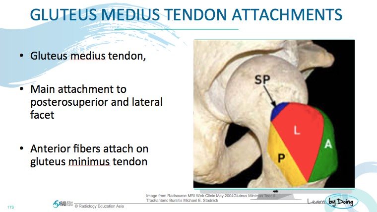
Image above: SP indicates Posterior superior facet where the main bulk of gluteus medium inserts.
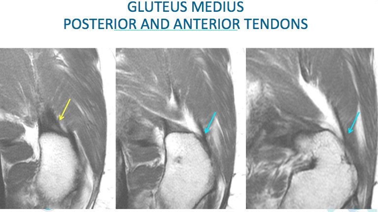
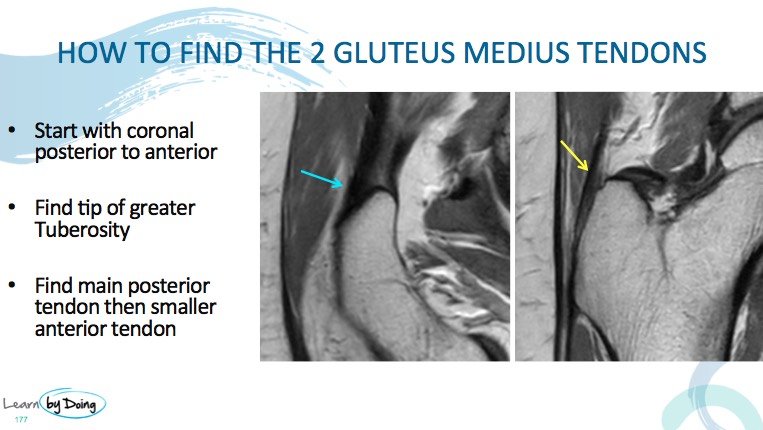
Image Above: Coronal scans posterior to anterior. Main bulk of gluteus medius fibres attach posteriorly to the tip of the trochanter.As you come anteriorly, fibres attach also to the lateral aspect of the trochanter.
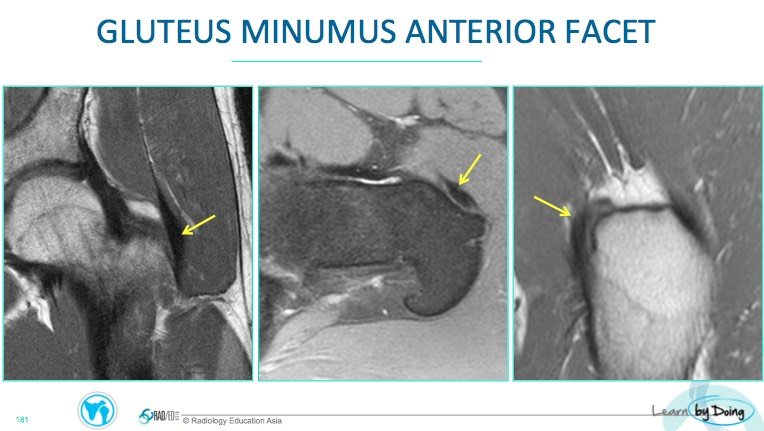
Image Above: Coronal, Axial and Sagittal scans of gluteus minimus attachment to anterior facet. Easiest to find on axial scans but make sure you can identify it on other planes.


