mri msk hip knee shoulder radiology education asia

MRI Hip How to Assess Acetabular Over Coverage

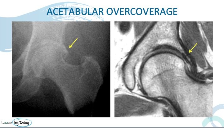
Acetabular over coverage which can be a cause of pincer type femoro acetabular impingement ( FAI) can be seen on MRI but is best assessed on xray. Review of the x-ray findings in acetabular overcoverage.

mri hip pincer fai acetabular coverage radiology education asia


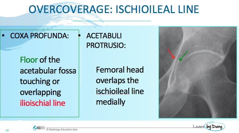
mri hip pincer fai acetabular coverage ischio ileal line radiology education asia

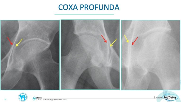
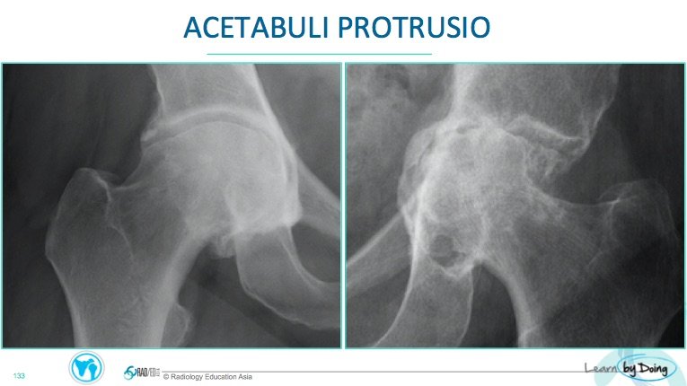
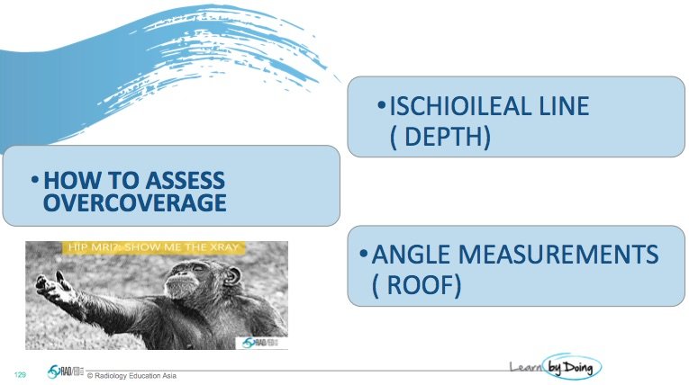
Image Above: Ischio Ileal line is is used to determine the depth of the acetabulum. Evidence of coxa profunda or acetabuli protrusion indicates over coverage.

mri hip pincer fai acetabular coverage ischio ileal line coxa profunda radiology education asiamri hip pincer fai acetabular coverage ischio ileal line acetabuli protrusio radiology education asia


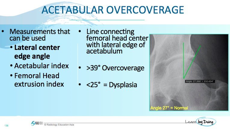
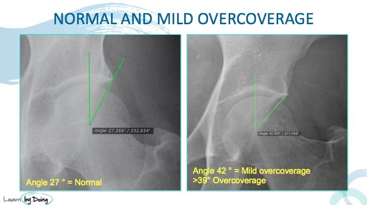
Lateral centre edge angle: Of the many measurements this is the easiest and will indicate if the acetabular roof is too large ( over coverage ) or is too small ( dysplastic)
mri hip pincer fai acetabular coverage lateral center edge angle radiology education asia

mri hip pincer fai acetabular coverage lateral center edge angle overcoverage radiology education asia

mri hip pincer fai acetabular coverage lateral center edge angle overcoverage mri radiology education asiaImage Above: Acetabular over coverage can be seen on MRI but x-ray is better and easier to assess.

Leave a Comment

Radiology Education

Join our Newsletter

Stay tuned on new
Mini-Fellowships launches and learnings

Mini-Fellowships

Better Than Conferences

We help radiologists learn by doing and report with confidence

Scroll to Top

Log in to your Mini-Fellowship or create an account

Mini-Fellowships

Better Than Conferences

We help radiologists
learn by doing and
report with confidence

Join our Newsletter

Stay tuned on new
Mini-Fellowships launches and learnings

Mini-Fellowships

Better Than Conferences

Join our WhatsApp Community

We help radiologists learn
by doing and report with confidence