MRI ELBOW LATERAL COLLATERAL LIGAMENT ANATOMY
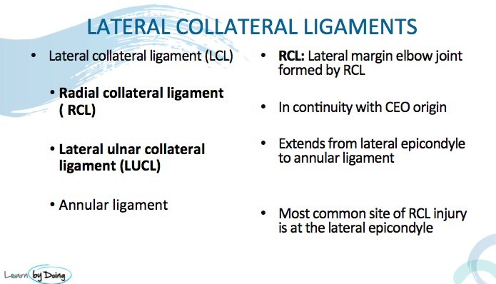
Ligament anatomy on MRI of the elbow is complicated but completely learnable if you are systematic about it. This post is on the Lateral Collateral Ligament (LCL) of the elbow. The LCL is composed of the Radial Collateral Ligament (RCL) and the confusingly termed Lateral Ulnar Collateral Ligament (LUCL). Again, its difficult to get a good understanding of the the MRI anatomy on static images but the aim of this post is to get a basic background understanding of the terms and locations so that when we look at the dicoms in the workshop, it won’t be totally foreign.
The two important things to know about the LCL.
- The Common Extensor tendon (CET) arises adjacent and proximal to the humeral attachment of the RCL. This means that a tear of the CET can extend into the RCL and we need to look at both structures when we assess the CET.
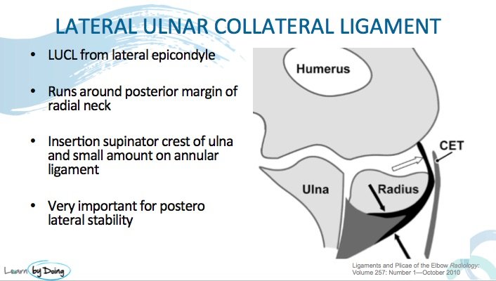
- The LUCL is important for postero lateral stability of the joint. A tear of the LUCL can lead to Postero Lateral Rotatory Instability ( PLRI).
Radial Collateral Ligament MRI Anatomy
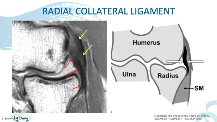
Image Above: RCL ( red arrows) forms the lateral joint margin and extends between the medial epicondyle and radius. Note the proximity of the Common Extensor Tendon attachment. Tears of the CET can extend into the RCL. White arrows in second image point to the RCL.
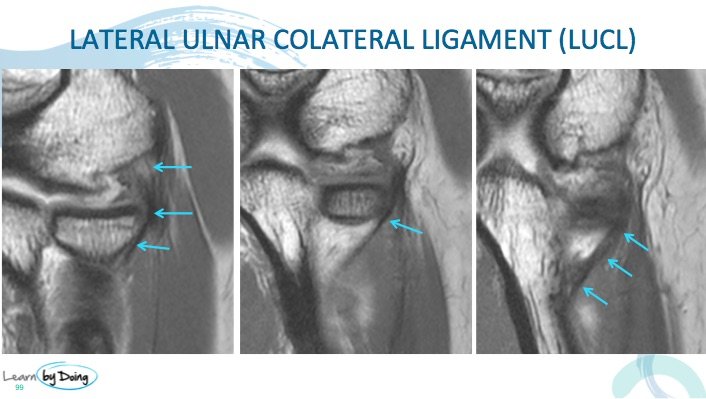
Image Above: LUCL ( blue arrows) extend from posterior aspect of medial epicondyle and sweep past the head and neck of the radius to insert onto the supinator crest of the ulnar. Note the striated appearance of the proximal attachment which is normal.

