MRI DENERVATION AROUND THE SHOULDER: PARSONAGE TURNER SYNDROME
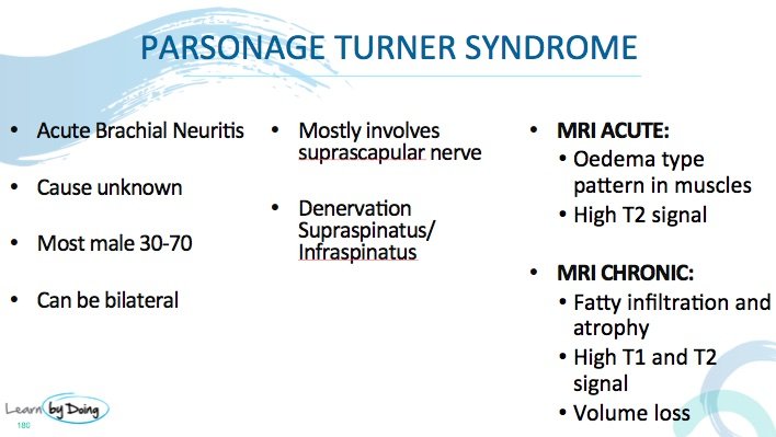
Parsonage Turner Syndrome is a brachial neuritis that can cause denervation of muscles around the shoulder joint. In the workshop we looked at the MRI features of Parsonage Turner Syndrome and in this post we have a quick review tof hose findings and also look at muscles other than supraspinatus and infraspinatus that can be involved. See also the previous post on MRI findings of denervation around the shoulder caused by paralabral cysts in the spinoglenoid and suprascapular notch at this link PARALABRAL CYST AND DENERVATION
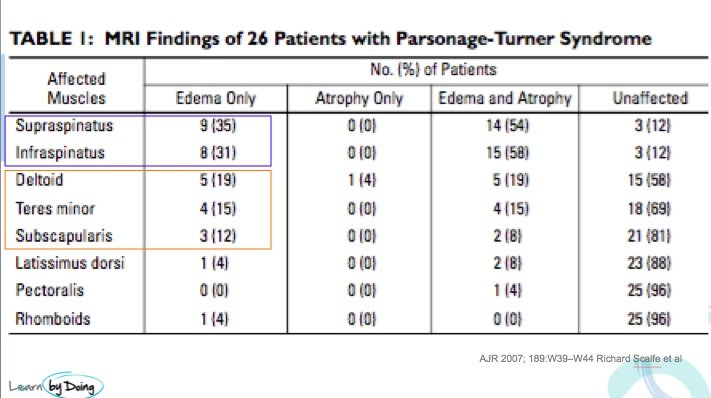
Image Above: Most common muscles involved in Parsonage Turner Syndrome are Supraspinatus and Infraspinatus. But there can also be involvement of deltoid, teres minor and subscapularis less commonly. From AJR 2007 189: W39-W44 Richard Scalfe et al.
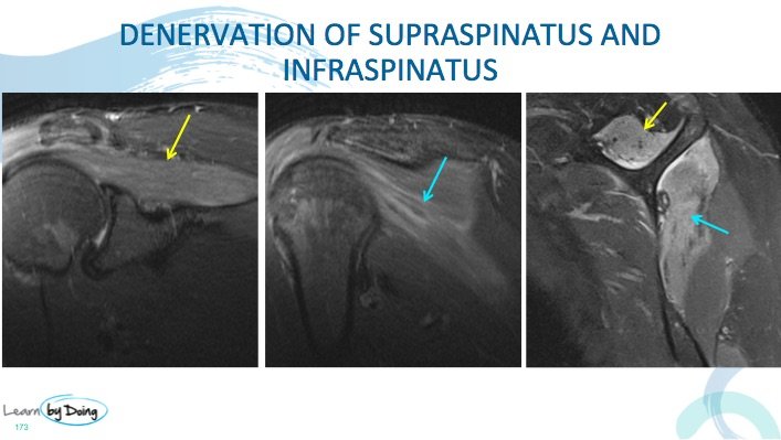 Image Above: Denervation of supraspinatus ( yellow arrow) and infraspinatus ( blue arrow). Increased signal on PDFS on keeping with denervation. No compression of suprascapular nerve seen.
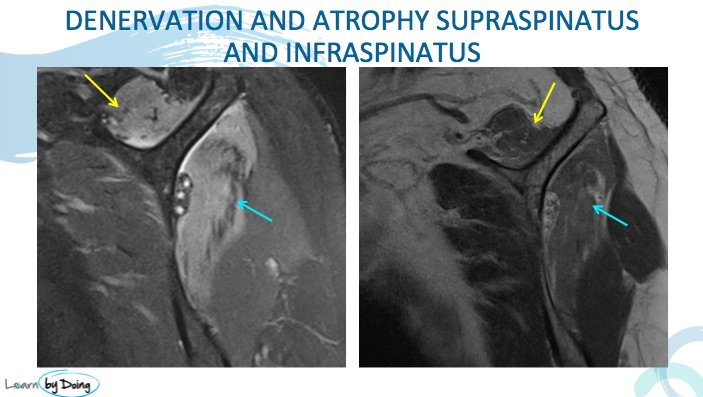 Image Above: Atrophy of supraspinatus (yellow arrow) and infraspinatus ( blue arrow). Loss of volume and fatty infiltration compare to normal teres minor ( not marked) just below infraspinatus. Compare PDFS and PD as non fat sat scan best to assess atrophy and fatty infiltration. High signal in denervated muscles on PDFS.
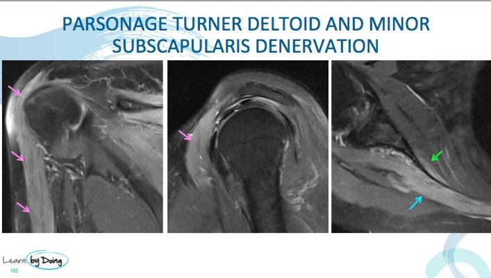 Image Above: Denervation of deltoid ( purple arrows) and very early subscapularis ( green arrow) denervation is less common but can be seen with Parsonage Turner Syndrome. Blue arrow infraspinatus denervation. No compressive structure seen.

