msk mri radiology conference

MRI ACL Mucoid Degeneration

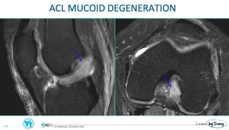
Mucoid Degeneration of the ACL on MRI can look similar to a tear. The cause is unknown but thought to be secondary to repetitive mictotrauma with micro tears and subsequent mucoid degeneration. This is what it looks like and how to differentiate it from a tear.

knee mri acl mucoid degeneration radiology education asia

knee mri acl mucoid microtear degeneration radiology education asia

Image Above: Hyperintensity and expansion of the ACL. ACL fibres are still seen in normal alignment within the hyperintensity.

knee mri acl mucoid degeneration tear radiology education asia

Image Above: Blue arrow ACL tear. ACL is hyperintense but no normal fibres seen. Yellow Arrow mucoid degeneration. ACL hyperintense and expanded and normal fibres see within it. Blue arrow cystic change which can be seen with mucoid degeneration.

knee mri acl mucoid degeneration edema oedema intraosseous cyst radiology education asia

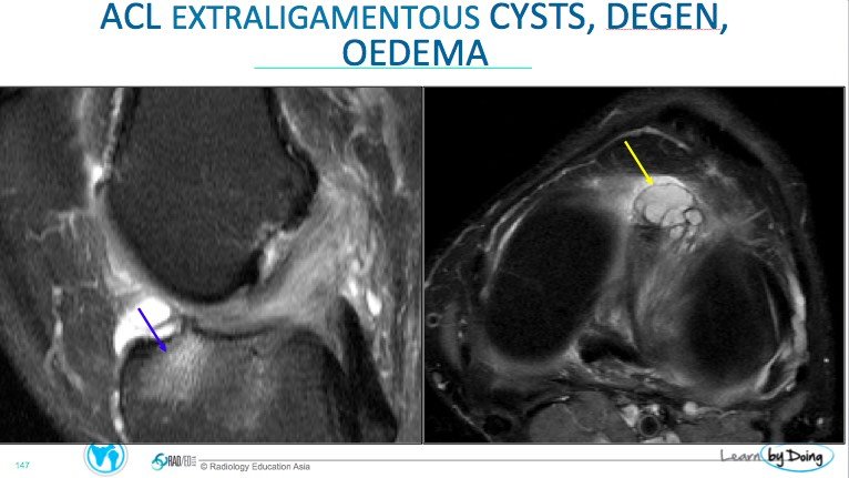
Image Above: ACL mucoid degeneration associated with bone marrow oedema ( blue arrow) and extra osseous cyst ( yellow arrow).

knee mri acl mucoid degeneration edema intraosseous cyst oedema radiology education asia

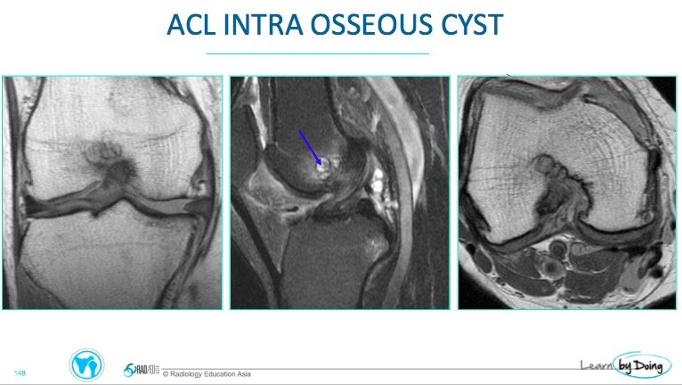
Image Above: Mild mucoid degeneration ACL associated with intraosseous cyst formation ( blue arrow).

Leave a Comment

Radiology Education

Join our Newsletter

Stay tuned on new
Mini-Fellowships launches and learnings

Mini-Fellowships

Better Than Conferences

We help radiologists learn by doing and report with confidence

Scroll to Top

Log in to your Mini-Fellowship or create an account

Mini-Fellowships

Better Than Conferences

We help radiologists
learn by doing and
report with confidence

Join our Newsletter

Stay tuned on new
Mini-Fellowships launches and learnings

Mini-Fellowships

Better Than Conferences

Join our WhatsApp Community

We help radiologists learn
by doing and report with confidence