Knee MRI MCL Tear
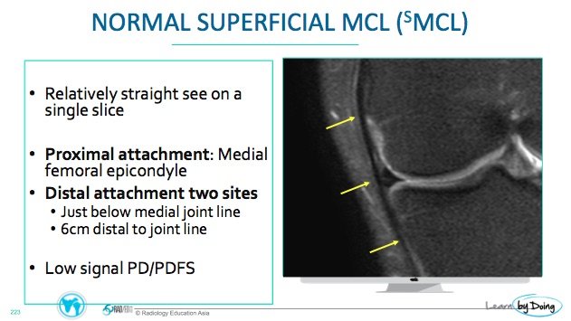
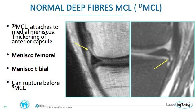
The medial collateral ligament of the knee (MCL) consists of the deep and superficial components. In this review we look at the appearance normal appearance of the MCL and of tears of the deep and superficial components on MRI.
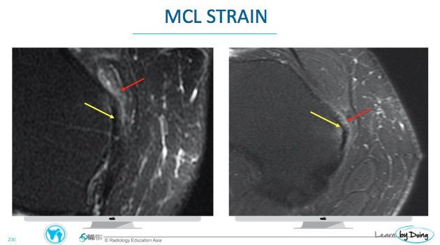 Image Above: Yellow arrow superficial MCL intact with oedema at the adjacent soft tissue ( red arrow) in keeoing with a strain.
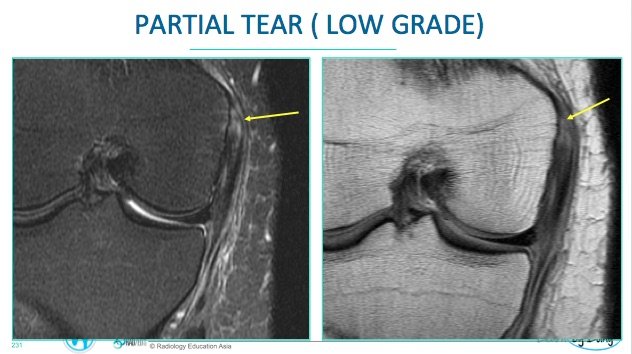 Image above: Low grade tear ( yellow arrow) woth localised loss of fibres and increased t2 signal.
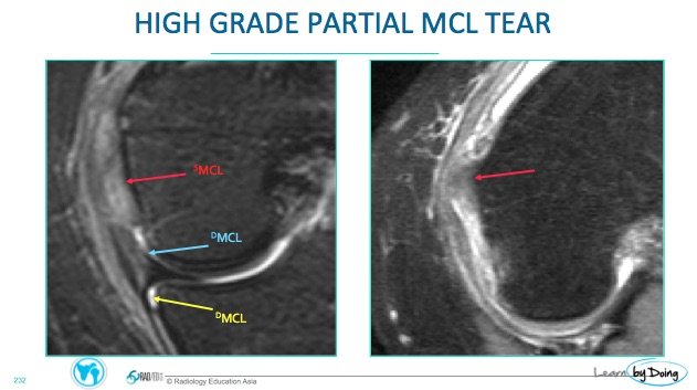 Image Above: High grade partial tear sMCL with absence of normal appearing fibres ( red arrow) but no retraction or loss of taughtness of sMCL. Tear of the meniscofemoral component of hte dMCL ( blue arrow). Meniscotibial dMCL intact ( yellow arrow).
 Image above: Complete tear sMCL. Loss of continuity of fibres and a wavy appearance indicates loos of attachment.

