ELBOW MRI ULNAR COLLATERAL LIGAMENT TEARS
In the last post we looked at the normal MRI anatomy of the Ulnar Collateral Ligament of the elbow and in this we look at tears. Get a sense of where to look and what to look for which will make it easier to understand and find when looking at the dicoms in the workshop.
Image Above: Tear and stripping ( blue arrows) of the distal ulnar attachment of the anterior band ulnar collateral ligament. The UCL should attach firmly to the ulnar ( at the sublime tubercle of the coronoid) and mostly the attachment is flush with the articular margin ( see first image in slide below). In this case there is stripping from the attachment with fluid signal seen between the ulnar and the UCL and thinning and high signal in the ligament ( centre image). Oedema is also present in adjacent flexor muscles.
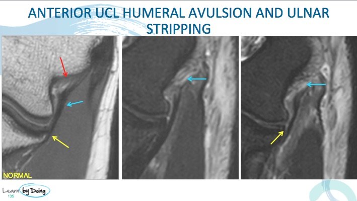
 Image Above: Tear ( blue arrows) of the proximal attachment of the anterior band ulnar collateral ligament. The first image is the normal striated appearance of the proximal attachment. Following two images demonstrate tear of the proximal attachment with oedema and laxity of the ligament. The ligament ( like all ligaments) should be taut and any laxity indicates rupture. Stripping also of the distal ulnar attachment ( yellow arrow).
 Image Above: Tear of the posterior band of the UCL ( blue arrows demonstrate normal and torn UCL). Often its easier to see on the axial images where it forms the base of the cubital tunnel. Look for the unlar nerve ( red arrow) and then go to the base of the tunnel to find a thin black line that forms the floor ( blue arrow) which is the UCL posterior band.


