ELBOW MRI COMMON EXTENSOR TENDINOSIS
The MRI appearance of tendinosis and tears of the Common Extensor Tendon is the same as for any other tendon and similar to the appearances we have seen in the shoulder rotator cuff. The main thing is to remember to also assess the Radial Collateral Ligament which we saw in the previous post arises immediately adjacent to the CET and can also be torn.
Image Above: Peritendinosis with fluid ( blue arrows) at the margins of the CET ( yellow arrows)
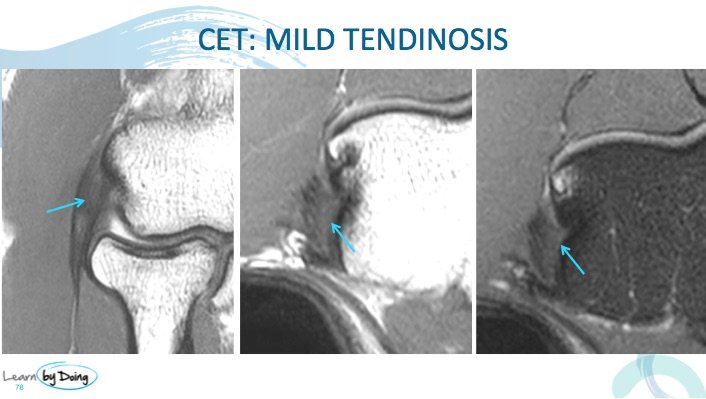
Image Above: Tendinosis common extensor tendon ( blue arrow). Increased PD and PDFS signal and expansion of tendon.
Image Above: Severe tendinosis common extensor tendon ( blue arrow). Increased PD and PDFS signal and expansion of tendon. Increased signal is not of fluid intensity to indicate a tear.
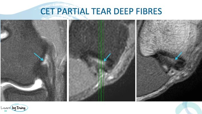
Image Above: Partial tear common extensor tendon ( blue arrow). Fluid intensity increased PD and PDFS signal.
 Image Above: Severe partial tear common extensor tendon ( blue arrow). Fluid intensity increased PD and PDFS signal.
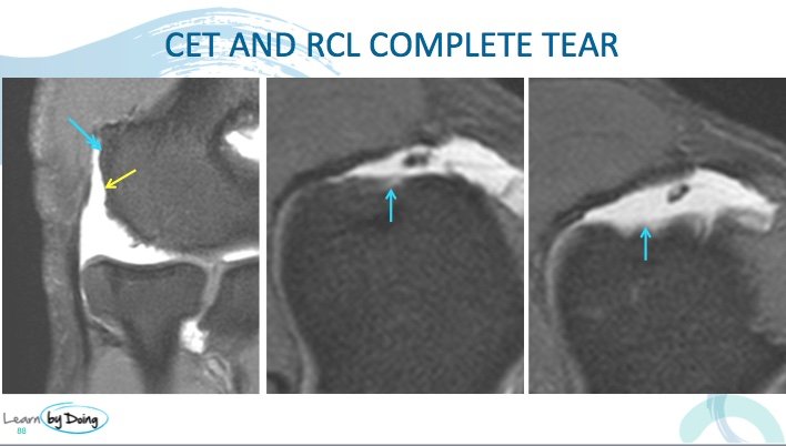
Image Above: Complete tear common extensor tendon ( blue arrow) and RCL ( yellow arrow). No normal fibres seen in their expected locations.


