ELBOW MRI BICEPS TENDINOSIS
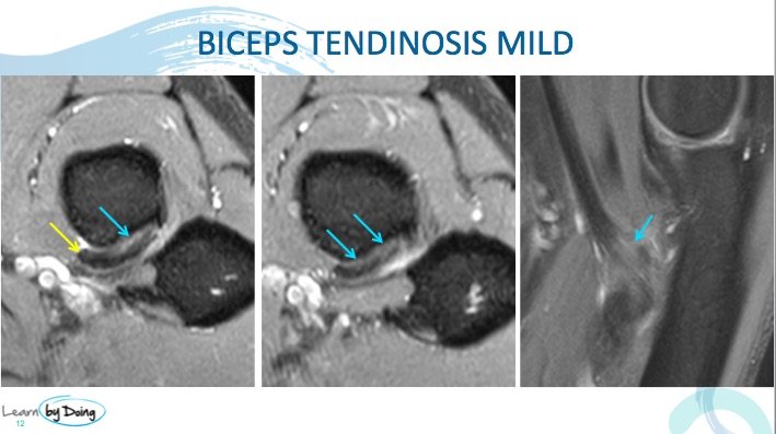
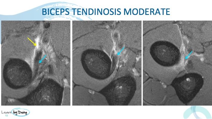
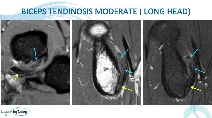
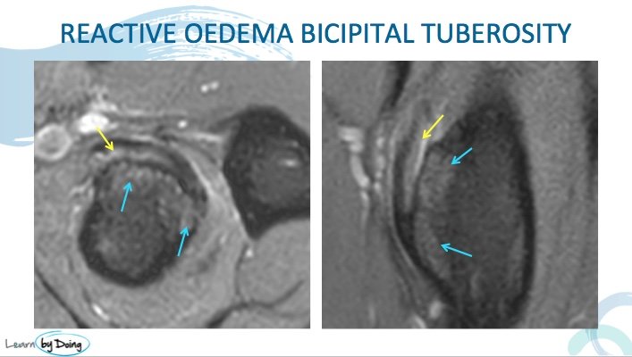
Biceps Tendon MRI. In this post we cover the MRI appearance of tendinosis. The changes seen are the same as we discussed in any other tendon with increased T2 signal, increased size and peri tendinous inflammatory changes. Tendinosis can be predominant in only one head of the biceps tendon and there can also be cystic changes or oedema at the bony site of attachment.
Image Above: Mild increased PDFS signal in tendon and peritendinous oedema ( blue arrow). Normal signal in proximal tendon ( yellow arrow).
hin fiImage Above: Thin fluid in bicipitoradial bursa ( yellow arrow) and moderate increased PDFS signal in tendon ( blue arrow).
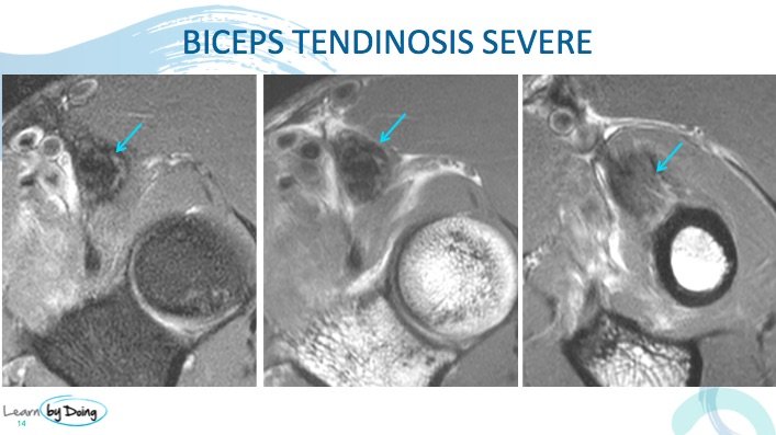
Image above: Severe tendinosis with ill definition, expansion and increased signal in tendon ( blue arrow).
Image above: Tendinosis affecting predominantly the long head of biceps tendon ( blue arrow). Short head which inserts more distally has mild tendinosis.
Image Above: Reactive oedema in the bicipital tuberosity ( blue arrow) and mild tendinosis biceps tendon ( yellow arrow).

