ELBOW MRI BICEPS TENDON ANATOMY
The biceps tendon inserts onto the bicipital tuberosity of the proximal radius and is composed of two tendons, the Short and Long Heads. Its always a bit difficult to get across MRI anatomy on static images and in the workshop we will be able scroll through the images and get a better sense of the anatomy. However its good to have a sense of what we will be looking at before we do so in the workshop.
Image above: Biceps tendon ( blue arrow) inserting onto the bicipital tuberosity ( yellow arrow).
Image Above: Long head of biceps ( purple tendon) inserts proximally and short head ( Blue Tendon) inserts more distally on the bicipital tuberosity. Dashed white region is the Bicipital Apponeurosis.
Image above: Images left to right proximal to distal. Long head of biceps tendon ( blue arrow) and short head ( yellow arrow) join to form a combined tendon ( red arrow).
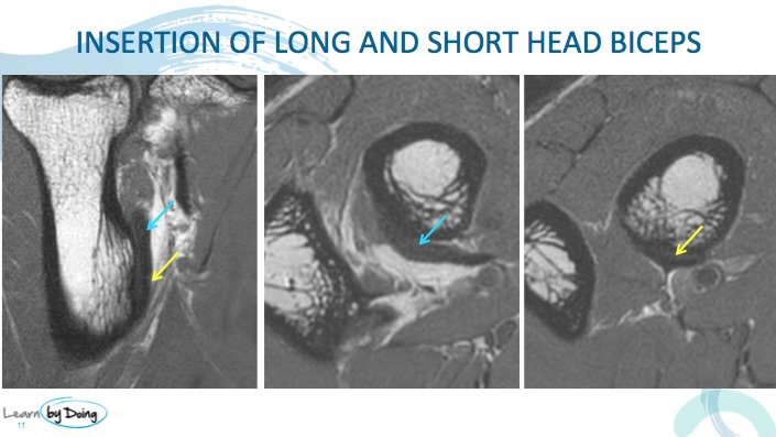
Image above: At insertion the long head fibres ( blue arrow) insert proximally and the short head ( yellow arrow) insert more distally.
Image above: White dashed region is lacertus fibrosis. Attached to biceps tendon and then fans out over proximal extensor muscles.
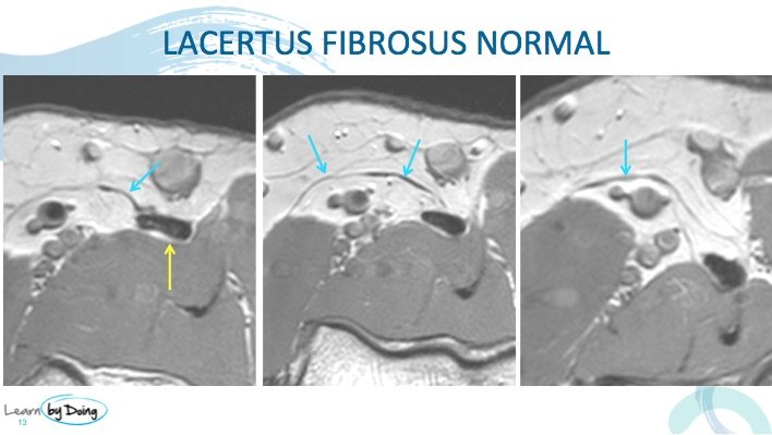
Image Above: Lacertus fibrosus ( blue arrow) arising from biceps tendon ( yellow arrow).
Image above: As annotated.

