CEREBELLAR INFARCTS: HOW NOT TO MISS THEM

The Problem: Acute cerebellar infarcts are easy to see on diffusion imaging, but small chronic infarcts in the cerebellum are often missed because they are confused with normal cerebellar sulci. How do you differentiate small chronic cerebellar infracts from normal sulci?

The Answer:
In the Cerebellar Hemispheres:

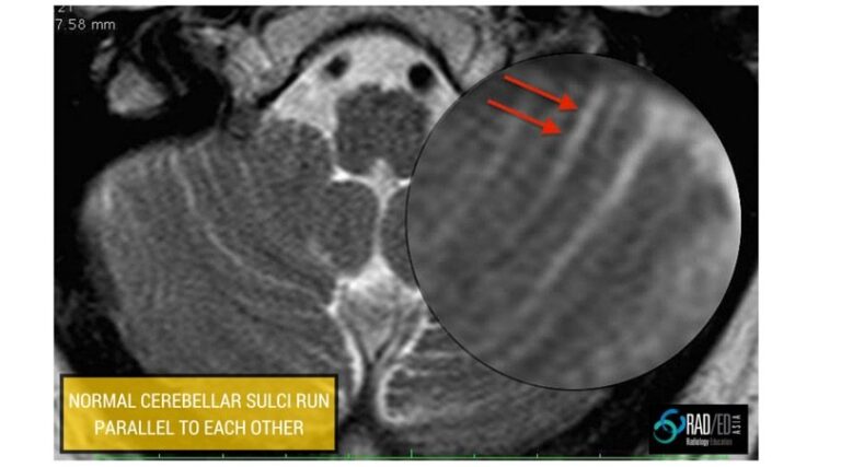
Normal:

cerebellar-infarcts-normal-sulci-mri

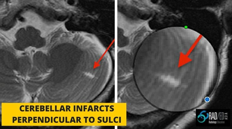
Chronic Infarct:

  • Infarcts usually run perpendicular to the sulci
  • If you see a CSF space running perpendicular to the sulcus its an infarct


cerebellar-infarcts-chronic-mri


In the Cerebellar Vermis:

Normal:

  • Both sides of the vermis should be symmetrical on axial and coronal images.

cerebellar-infarcts-vermis-mri-normal


Chronic Infarct:

  • If its assymetric with parts of the vermis missing that is a sign of infarction.

Leave a Comment

Radiology Education

Join our Newsletter

Stay tuned on new
Mini-Fellowships launches and learnings

Mini-Fellowships

Better Than Conferences

We help radiologists learn by doing and report with confidence

Scroll to Top

Log in to your Mini-Fellowship or create an account

Mini-Fellowships

Better Than Conferences

We help radiologists
learn by doing and
report with confidence

Join our Newsletter

Stay tuned on new
Mini-Fellowships launches and learnings

Mini-Fellowships

Better Than Conferences

Join our WhatsApp Community

We help radiologists learn
by doing and report with confidence