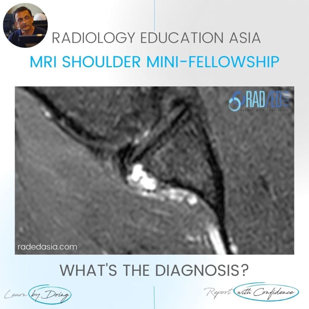
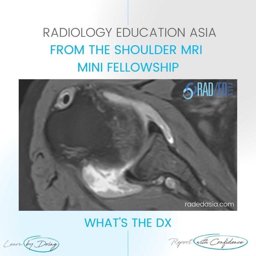
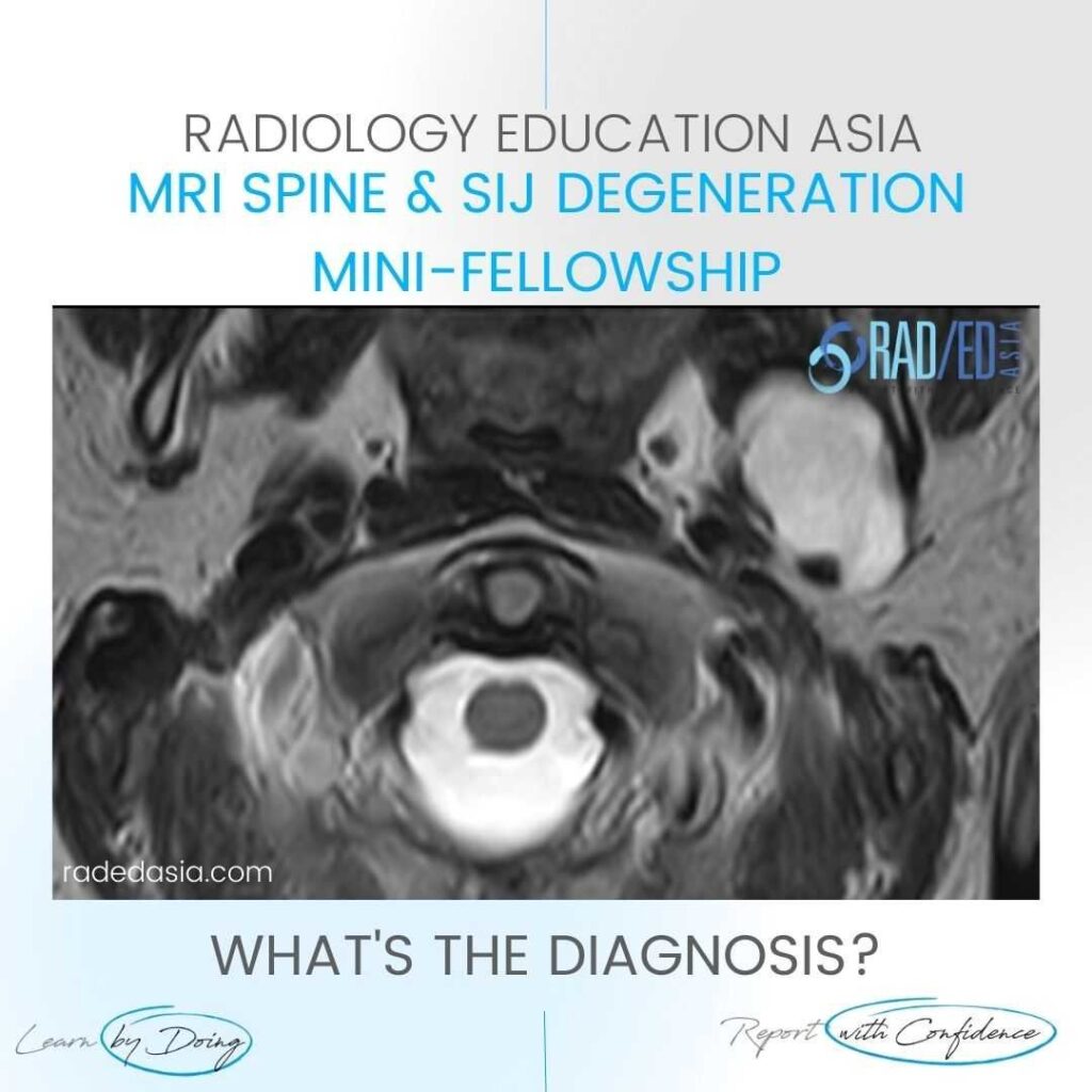
SHOULDER PARALABRAL CYST WHAT ARE THE FINDINGS? High signal cystic structure (Pink arrow) adjacent to the labrum. DISCUSSION: WHAT'S THE Dx? The structure is a paralabral cyst (Pink arrow) lying adjacent to the posterior labrum. Paralabral cysts are usually seen adjacent to the labrum but can occasionally track away from the labrum. They […]
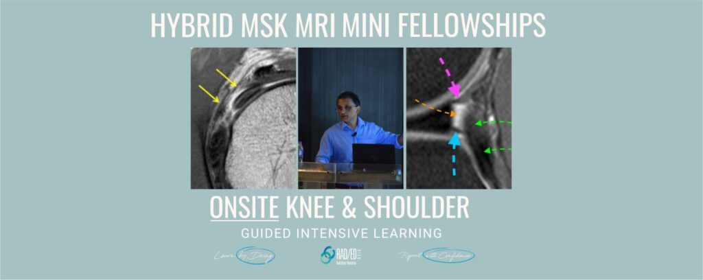
Home MSK RADIOLOGY CONFERENCE COURSE MUSCULOSKELETAL MRI INDIA NEXT ONSITE MSK MRI RADIOLOGY WORKSHOP CONFERENCE: 17 & 18 SEPTEMBER 2022 Our new onsite Hybrid MSK Musculoskeletal MRI Conference and Course in India will build on our Online MSK MRI courses with: More advanced topics not covered in the Online courses. Guest lectures on new and
BICEPS TENDON DISLOCATION RADIOLOGY MRI BICEPS TENDON DISLOCATION ON MRI: We normally look for Biceps dislocation on axial scans, but you can see it as well on Coronals. In this Quick video we look at: The Normal location of the Biceps tendon. What happens to the tendon on coronal scans when it dislocates.
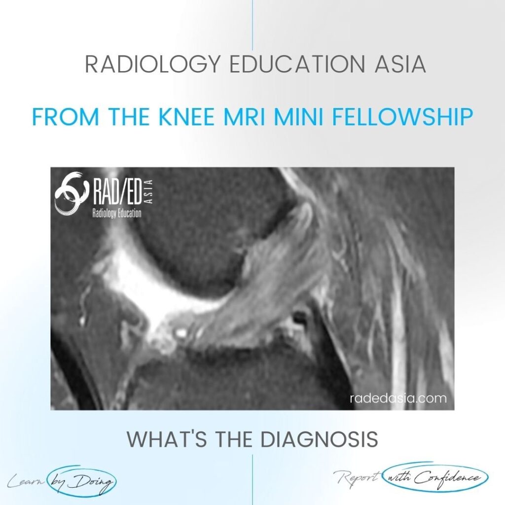
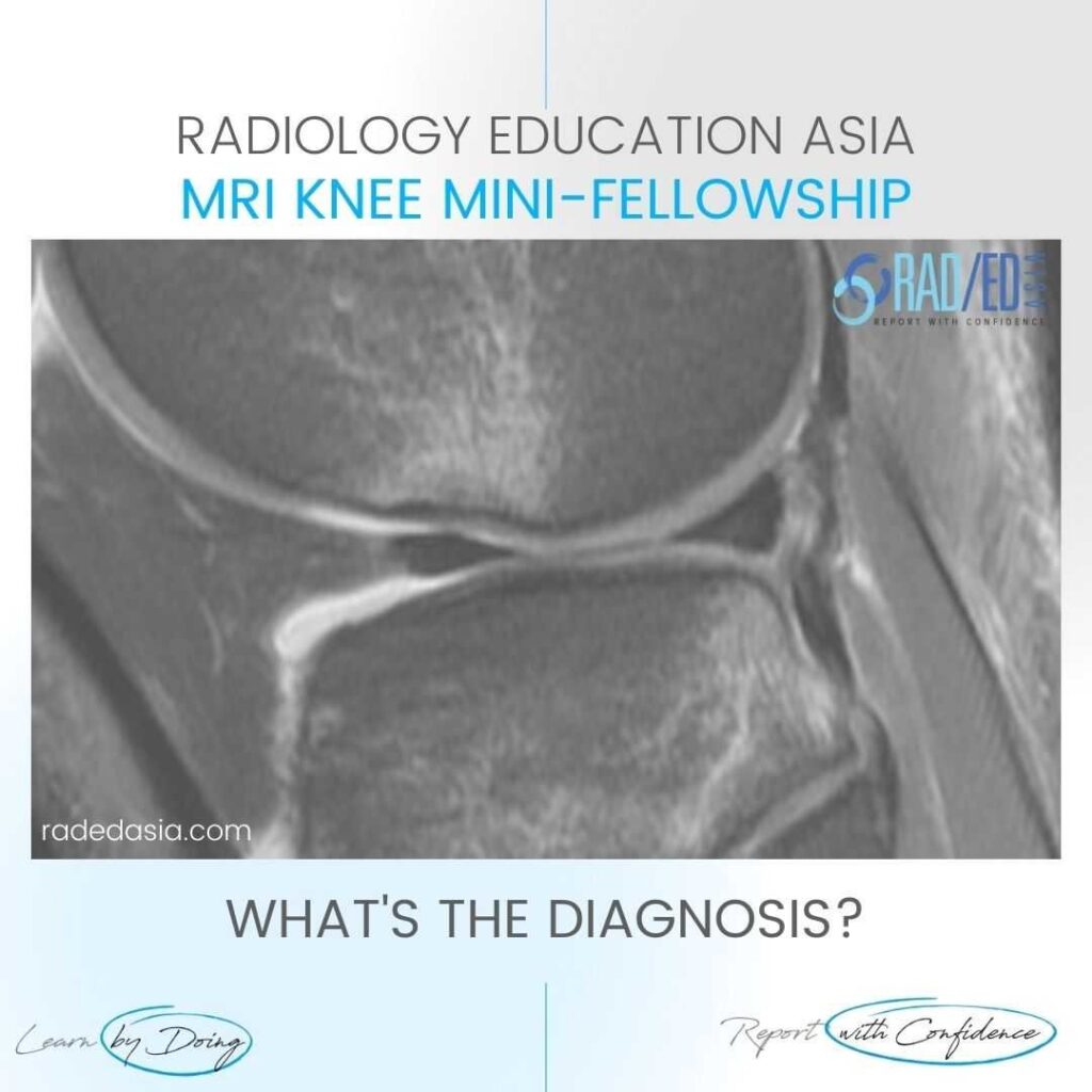
ACL MUCOID DEGENERATION RADIOLOGY MRI DISCUSSION Mucoid Degeneration. These findings indicate Mucoid Degeneration of the ACL rather than a tear. This is a characteristic appearance of ACL mucoid degeneration where the ACL is expanded and hyperintense but is still continuous with normal alignment. . WHAT ARE THE FINDINGS The ACL is enlarged and hyperintense
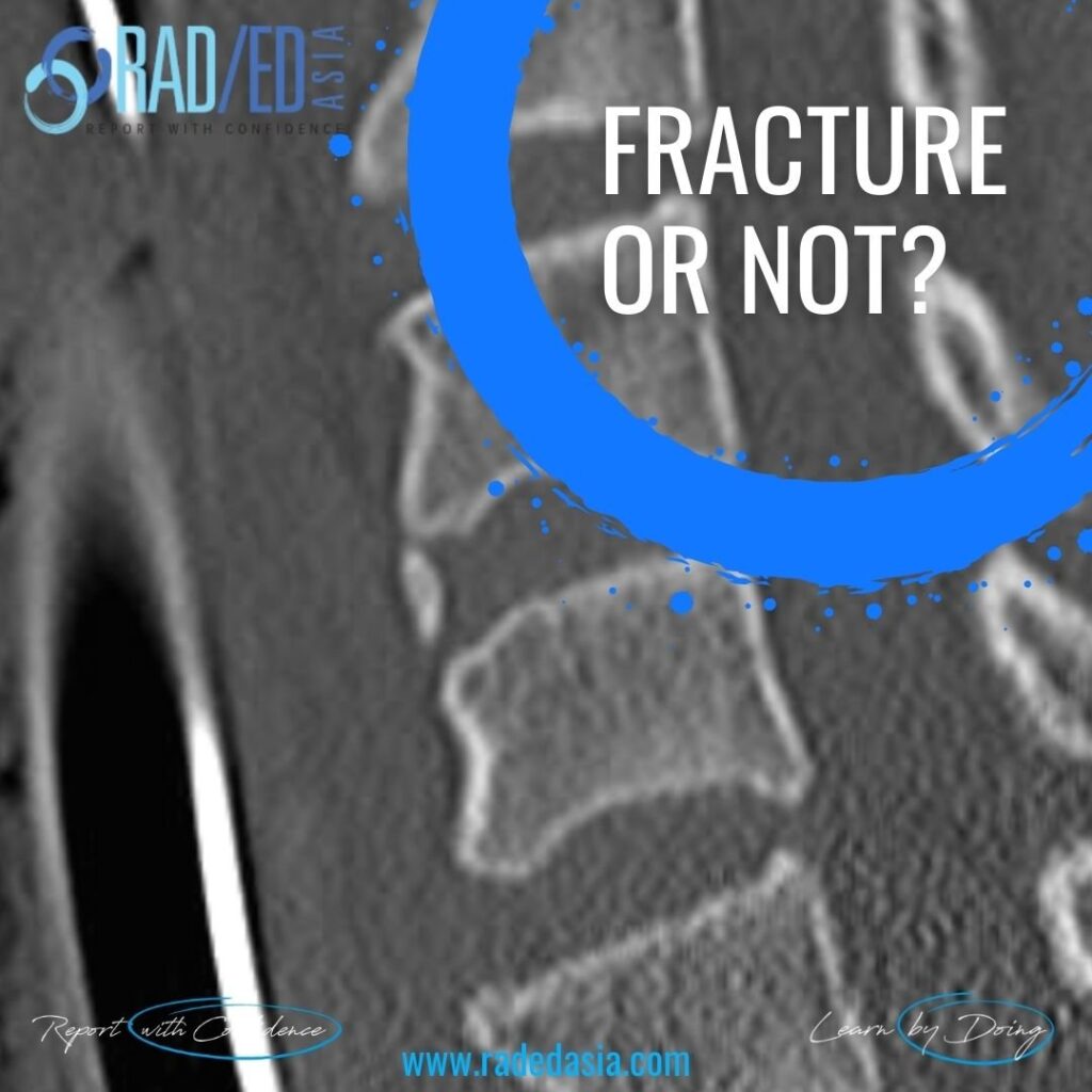
CERVICAL TEARDROP FRACTURE OR NOT IN TRAUMA RADIOLOGY HISTORY The patient has had trauma with a cervical fracture seen elsewhere. Is this a teardrop fracture? What do you think? VIEW VIDEO FINDINGS This is not a teardrop fracture but chronic ossification in the annulus. The reasons why this is not a fracture is that
SPINE AND CORD TRAUMA IMAGING RADIOLOGY COURSE X-RAY CT MRI SPINE AND CORD TRAUMA IMAGING RADIOLOGY: What’s in the New Spine and Cord Trauma Imaging course? A quick video explaining all that we have to help you assess and report Spinal and Cord Trauma more confidently. https://vimeo.com/713147494 #radedasia #radiology #radiologist #radiologia #spinetrauma #emergencymedicine #emergencyradiology
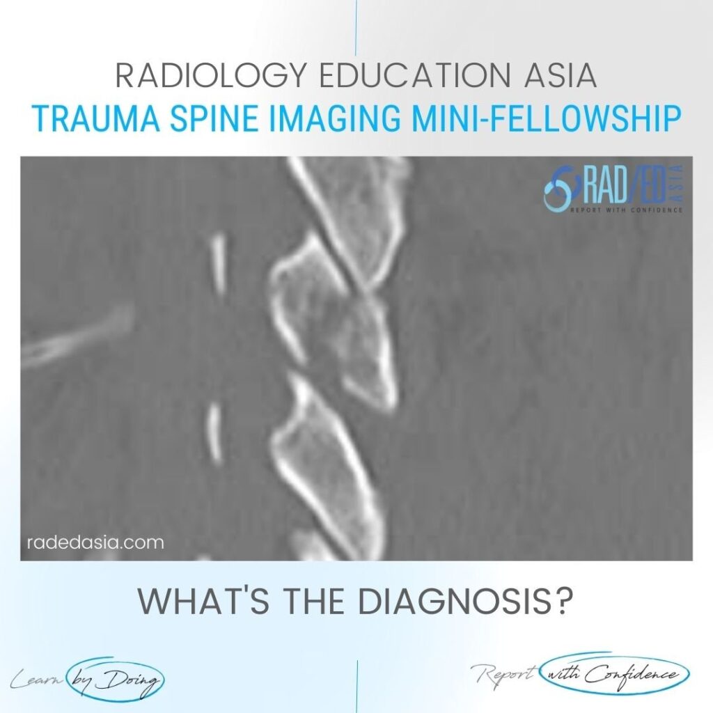
SPINE TRAUMA RADIOLOGY: FACET DISLOCATION ON CT FACET DISLOCATION ON CT: Spinal trauma can result in a number of abnormalities of the facet joints ranging from Diastasis, Subluxation, Perched Facets and eventually to Dislocation or Locked Facets. In this short video we look at the normal appearance of the facet joints, how that
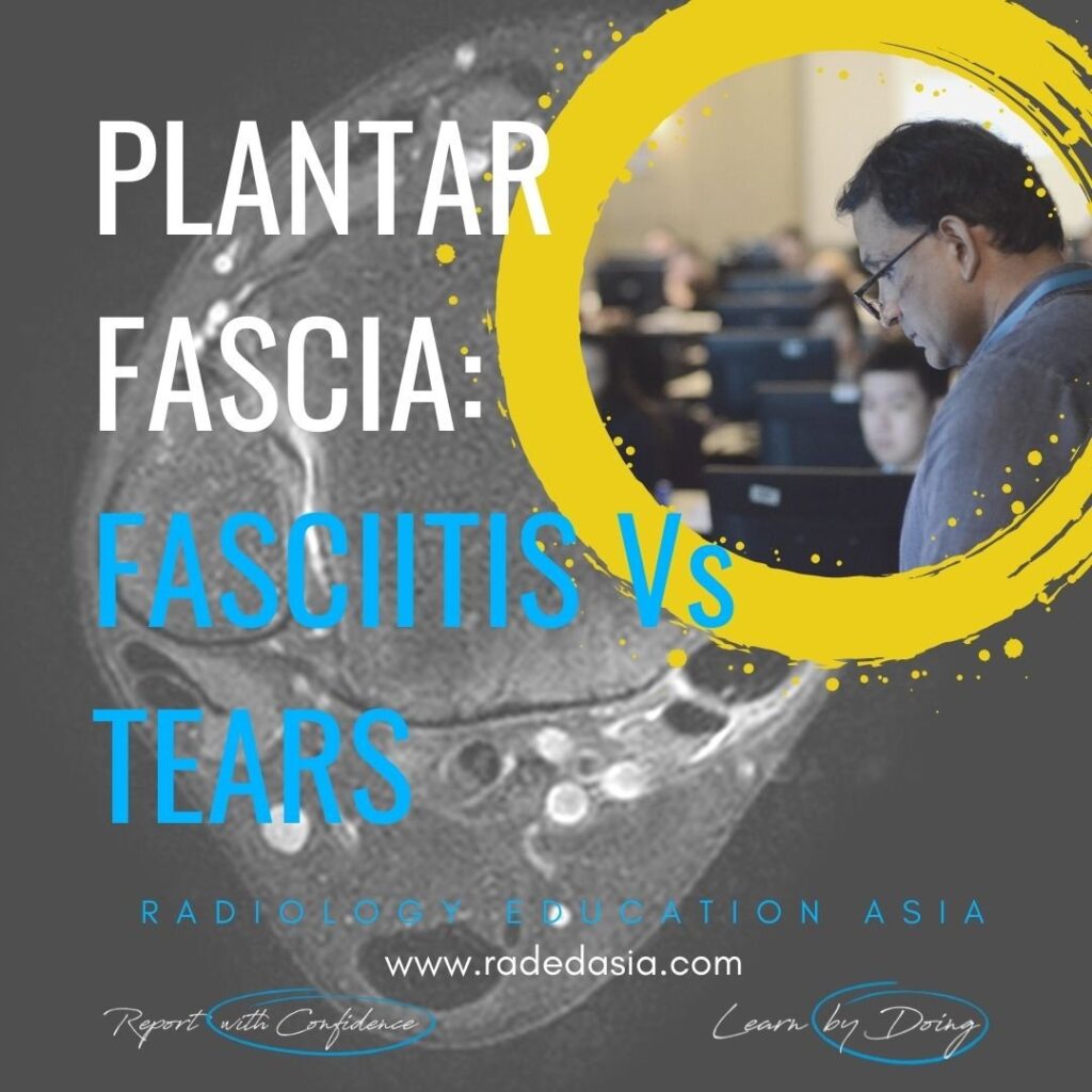
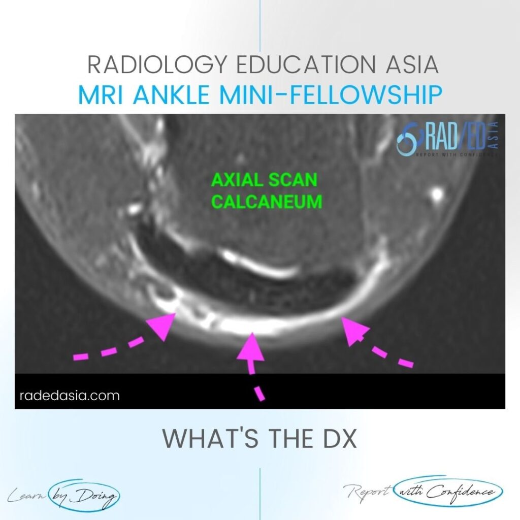
PLANTAR FASCIA TEAR vs PLANTAR FASCIITIS HOW TO DIFFERENTIATE BETWEEN PLANTAR FASCIA TEAR vs PLANTAR FASCIITIS: How do you tell the difference on MRI between Plantar Fasciitis and Plantar Fascia Tears. Short 1 minute video on the key differences that help you differentiate a Plantar Fascia Tear from Plantar Fasciitis on MRI. VIEW VIDEO
THE ANSWER TO MEDICAL BURNOUT… KOMODO DRAGONS The Answer to medical burnout… No matter how interested you are in your work and how interesting the work is, I think everyone gets to a point where it starts to become routine and you start looking for something to light up the day. That’s what was happening
RETROACHILLES BURSA BURSITIS MRI ANKLE DISCUSSION ABOUT THE RETROACHILLES BURSA The retroachilles bursa lies between skin and Achilles tendon. It’s not usually seen as its an adventitial bursa caused by chronic friction. It is Uncommonly enlarged. WHERE TO LOOK FOR THE RETROACHILLES BURSA Best seen on axial and sagittal scans. Look posterior
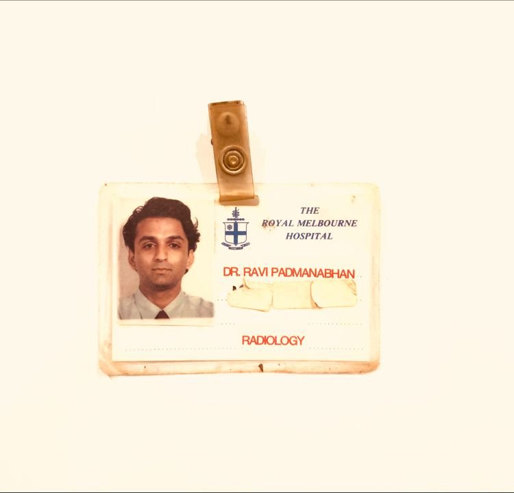
THE BENEFIT OF REJECTION How did I end up choosing MRI as my sub specialty? The benefit of Rejection. I had just finished my Radiology training from Royal Melbourne Hospital. It was time for the final step, the fellowship. I had always been interested in Ultrasound and particularly Obstetric Ultrasound but my own college didn’t
THE BENEFIT OF BEING OVERWORKED Why Radiology? The benefit of being Overworked. After finishing medicine at the University of Melbourne, I was intent on being a Physician and was in the Physicians training programme. It may have changed now, but the days were overwhelming, with very long and exhausting shifts. It wasn’t uncommon to start
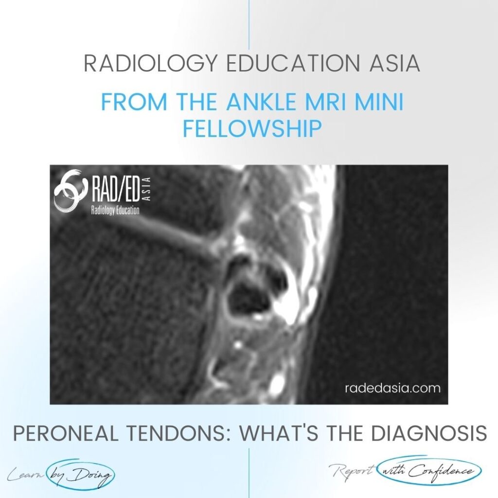
PERONEUS BREVIS TENDON TEAR MRI DISCUSSION: WHAT'S THE dX Peroneus Brevis Tendon Tear: MRI demonstrates a peroneus brevis tendon tear. There is a longitudinal split resulting in two components of the Peroneus Brevis tendon (Pink arrows). Peroneus Longus (Blue arrow) is normal. WHAT ARE THE FINDINGS There should only be two tendons in the peroneal
SHOULDER SYNOVITIS MRI RHEUMATOID ARTHRITIS DISCUSSION: WHAT'S THE Dx? Extensive synovitis and ACJ erosions from Rheumatoid arthritis. Having synovitis in multiple locations (even if there are no erosions) should raise the possibility of an inflammatory abnormality. Thank you to Dr Joe Thomas (Rheumatologist) for the case. WHAT ARE THE FINDINGS? Image 1: The first
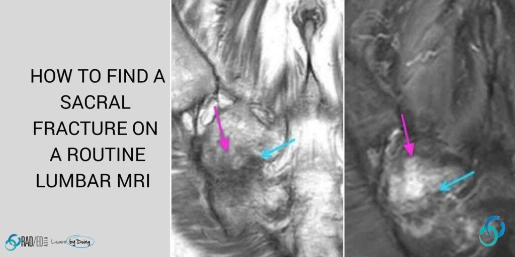
SACRAL FRACTURE MRI: HOW TO FIND ON A LUMBAR MRI HOW TO FIND SACRAL FRACTURE ON LUMBAR MRI When the clinical examination and referral notes are inadequate (Back Pain F.I.) the cause of the pain may well be sacral rather than lumbar. On a standard lumbar spine MRI with axial and sagittal scans, the sacrum
ACL TEAR PIVOT SHIFT INJURY BONE CONTUSION PATTERN OSTEOCHONDRAL FRACTURE MRI WHAT'S THE Dx? Pivot shift bone bruising of the lateral femoral condyle and an osteochondral depression fracture secondary to an ACL tear. Characteristic bone contusion pattern seen with complete ACL tear. The osteochondral fracture is seen with the flattening of the cortical
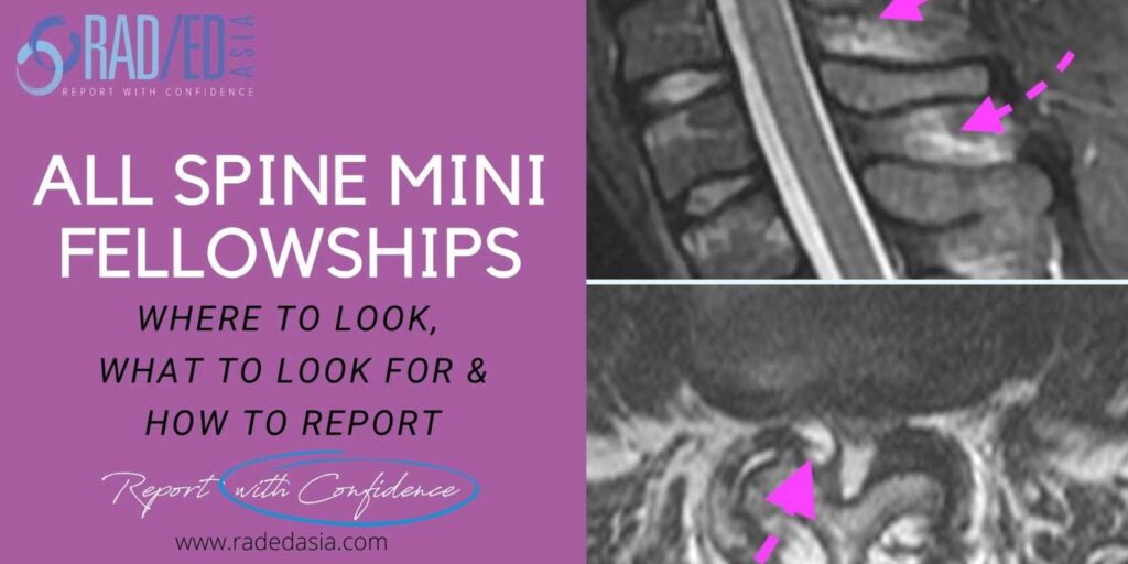
Home SPINE IMAGING COURSE: SPINE MRI CT XRAY RADIOLOGY Spine Imaging Courses. Learn about imaging the spine in our Guided Spine Imaging Mini Fellowships. 30 Day courses that cover the Anatomy, Pathology that assists in the diagnosis and Imaging of spinal abnormalities. Where to Look, What to Look for and How to Report…With Confidence. OUR
FACET FRACTURE RADIOLOGY CT DISCUSSION FACET FRACTURE CT APPEARANCE The facet is fractured (Pink arrows) but there is no evidence on this image of facet subluxation or dislocation. However the facet joint is widened inferiorly (Green arrow) indicating that the facet capsule must be torn. FACET FRACTURE RADIOLOGY CT: SLIDER TO VIEW IMAGES ARE
