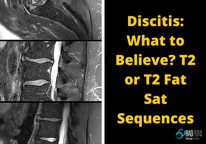
When scans are performed for ? Discitis, often both a T2 and T2 Fat Sat/ STIR sequence are performed. Which is better for assessing for early discitis? One of the features of discitis on MRI is increased T2 signal in the disc. But often, there are no other features of discitis/ osteomyelits, the disc signal […]
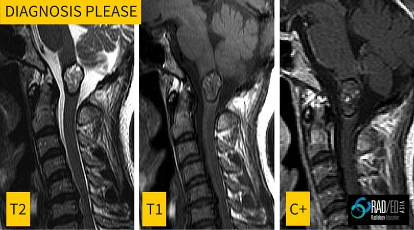
This is the first in a regular series of posts on cases of interest. Incidental finding in a 40yo male. Image above. ANSWER Cavernomas of the spinal cord have a very specific appearance on MRI with the following features Lobulated well defined lesion mixed signal T1 and T2 No significant enhancement Low signal
In the last post we looked at the Buford Complex. In this post lets look at the remaining two normal labral variants, the Sublabral Recess and the Sublabral Foramen Definition: Regions where a normal labrum is present but it is not attached to the glenoid. Where are they found? Sublabral Recess: Anterior portion of the
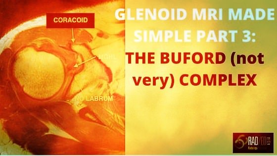
In this third part of Glenoid Labrum MRI Simplified we look at the Buford Complex. If you have not seen Part 1 and Part 2 please look at those posts before reading this. Definition: Absent anterosuperior labrum, with a thick middle glenohumeral ligament. MRI APPEARANCE The image below shows a comparison of the normal appearance
The problem: Many normal labral variants get called tears. How do we differentiate a normal labral variant from a tear? The solution: Normal labral variants occur in specific areas of the glenoid labrum. Understand their typical location and you will not confuse them with tears. If you havent read the first post on the classification
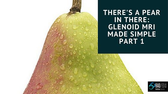
The problem: A lot of the difficulty people have in reporting shoulder MRI and particularly the glenoid labrum is in having a clear understanding of the MRI anatomy. This leads to confusion in differentiating normal glenoid labrum variants from labral tears and SLAP tears. The solution: The humble pear. Only a few dollars a kilo
PARS DEFECTS ON MRI Pars defects can be difficult to see on MRI. Lets look at how best to find them 1. WHAT IS THE PARS INTERARTICULARIS Bone that connects the superior and inferior articular process at each level. 2. WHAT DOES A NORMAL PARS INTERTICULARIS LOOK LIKE ON MRI A continuous piece of bone
INTRO: Pars defects can be difficult to see on MRI. Here is an easy way to know if the slip/ spondylolistheisis you are seeing is due to a pars defect or is from degenerative facet disease. THE TIP: In Degenerative spondylolisthesis, the canal is always small. In spondylolisthesis due to a Pars Defect, the
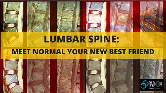
In our workshops, before we look at abnormal, we always look at the normal appearance of a structure and make sure we understand it well. Without knowing normal, you can’t hope to understand what is abnormal. So what does a normal lumbar disc look like on MRI and what is the normal degenerative process of
In our hands on workstation imaging workshops, we are often asked which Dicom viewers we recommend. Here is the simple answer. For Mac there are two options: Osirix: Is the best known Dicom Viewer which I have used for many years. It stores, anonymises, organises
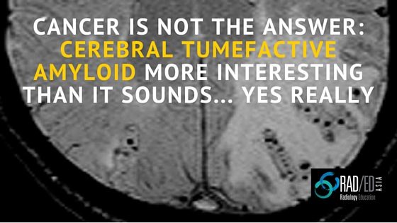
INTRO: Cerebral tumefactive amyloid is a rare mass like form of cerebral amyloid. PATHOLOGY: Caused by vasculitis +/- perivasculitis which is an autoimmune response to the amyloid deposition. KEY IMAGING POINTS: MRI findings similar to a glioma/ treated lymphoma with T2 hyper intensity and mass effect. Can demonstrate leptomeningeal enhancement over the region of abnormality, however
RadEdAsia ( Radiology Education Asia) has been providing hands-on workshops in MRI in the Asia/ Australia region for over 10 years. We have concentrated on MRI education and our old website was fairly basic. But all that is now changing. Beginning 2016 we have expanded the list of MRI workshops and we will also be providing
This was the first time that I have been to the National Neuroscience Institute’s (NNI) Advanced Neuroradiology Course despite living in Singapore for over 10 years. I tend to limit the number of conferences I attend as I find that the practical educational return for the amount of money and time spent is poor. I
Quick Journal Review: Should you use T2* or SWI sequences to detect cerebral microbleeds.Which sequence is more sensitive? Journal Article: SWI or T2*: Which MRI Sequence to Use in the Detection of Cerebral Microbleeds? The Karolinska Imaging Dementia Study S. Shams et al AJNR 36:1089–95 Jun 2015 Intro: Different sequences can be used to detect
Most radiology articles, books, websites and conferences are in English. Great if you speak and understand English well, but in the 10 years of giving talks and hands on workshops in the SE Asian region, I have found that most people would prefer the information,and will learn a lot more, in their own language. So
