CLICK HERE FOR OUR LATEST SPINE MRI MINI-FELLOWSHIPS Radiology Conference Malaysia Kuala Lumpur. This meeting is now over. If you would like to see photos from the event, please follow this link to our Facebook page KLSpinePhotos We have had a lot of requests to hold the Spine Workstation Workshop in Kuala Lumpur as well […]
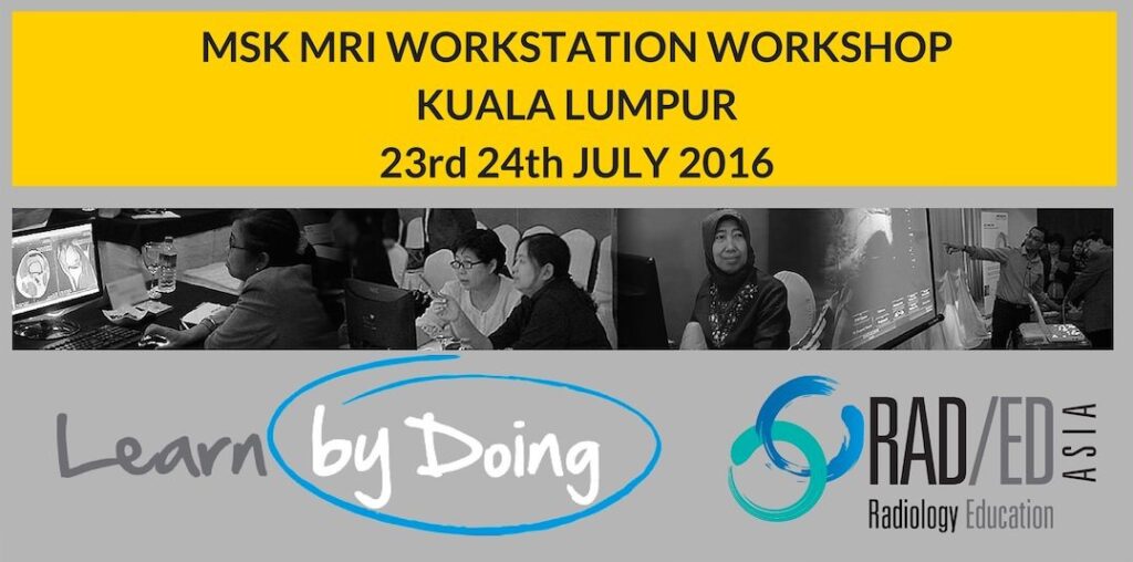
CLICK HERE FOR OUR LATEST SPINE MRI MINI-FELLOWSHIPS Radiology Conference Malaysia Kuala Lumpur MSK MRI This workshop is now over. We had a great response to the workshop and will be shortly announcing dates for the last two workshops in KL this year which will be MRI Upper Limb and Spine MRI. If you would
CLICK HERE FOR OUR LATEST SPINE MRI MINI-FELLOWSHIPS This Meeting is now over. Links below to our next workshops Spine and SIJ MRI Workstation Workshop Kuala Lumpur 13th November 2016 MSK MRI Workstation Workshop Kuala Lumpur Malaysia 11-12 February 2017 We will be holding a 1 day CT/MRI workstation based workshop in Nepal, on the
We will be holding a 1 day CT/MRI workstation based workshop in the Phillipines in Cebu, on the 16th of July 2016. We will be covering CT assessment of the Lower Limbs and SI Joints and MRI of the Shoulder. This is a private event for Philippines radiologists who have already been invited and unfortunately
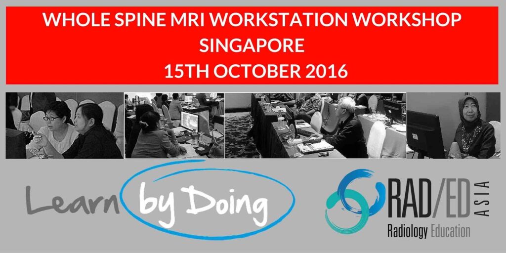
CLICK HERE FOR OUR LATEST SPINE MRI MINI-FELLOWSHIPS We will be running a one day intensive MRI Spine Workstation Workshop in Singapore on Saturday 15th of October. This will cover all aspects of Spine and Sacro Iliac Joint imaging. Like all of our workshops, this will be individual workstation based with guided viewing of hundreds
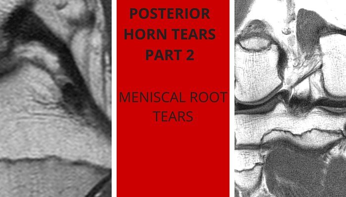
What is the meniscal root? The meniscal root is a small portion of the meniscus that attaches and anchors the anterior and posterior horns of the meniscus to the intercondylar fossa or eminence of the tibia. Its usually the posterior root of the meniscus that is torn and medial more than lateral meniscus. Where is
MSK MRI Workstation workshop Delhi India October the 1st and 2nd 2016 will be covering MRI of the Knee, Shoulder, Hip and Ankle in an intensive, structured learning, workshop over two days with lectures and guided viewing of 100’s of full Dicom studies of MRI pathology on individual workstations, so that you learn and apply
POSTERIOR HORN MENISCUS: Three posts for such a small thing. How can it be so important? ANATOMY: The posterior horn has two components 1. The posterior horn itself 2. The meniscal root which attaches the posterior horn to the intercondylar fossa TYPES OF TEARS: The posterior horn can have the usual types of meniscus tears
When was the last time you saw a football or cricket match where the players were messaging or checking their Facebook page in between playing? The thought of a sportsperson pulling out a phone to check a Whats app message or Facebook status update seems ridiculous. Why? Because its obvious that checking your phone in
I catch a lot of taxis and anyone who has experienced the taxi system in Singapore knows how fantastic it is. But what makes it like that? Taxis are plentiful and affordable Taxis are modern, clean, safe and comfortable. Honesty: I have yet to be cheated or more importantly, even feel that I might have
We had a great response to the MSK MRI Workstation workshop to be held in July in Kuala Lumpur and had to stop registrations within a week. There have been a lot of further enquiries and because learning is better in smaller groups, rather than have more registrants in the July session,we have decided to
The Problem: Acute cerebellar infarcts are easy to see on diffusion imaging, but small chronic infarcts in the cerebellum are often missed because they are confused with normal cerebellar sulci. How do you differentiate small chronic cerebellar infracts from normal sulci? The Answer: In the Cerebellar Hemispheres: Normal: Chronic Infarct: Infarcts usually run perpendicular to the
Intro: With an anterior dislocation, the anterior glenoid can be damaged by the humeral head as it dislocates. Acute Dislocation: This can be an acute fracture with avulsion of the fragment and acutely you will see bone marrow oedema in the glenoid which alerts you to the injury and makes it easy. Chronic Dislocation: But
CHONDRAL DELAMINATION CARTILAGE MRI CHONDRAL DELAMINATION CARTILAGE MRI Chondral Delamination on MRI is not common but is seen often enough that we need to be aware of it and What to look for on MRI. OVERVIEW What is it? Chondral delamination is when cartilage separates and lifts off from its attachment with the cortex.
Registrations have now closed for the MSK MRI Workstation based workshop to be held in Kuala Lumpur Malaysia in July 2016. We would like to thank everyone who registered and to those who expressed interest. Due to continuing interest in the workshop we have decided to hold a second workshop in KL on the weekend
