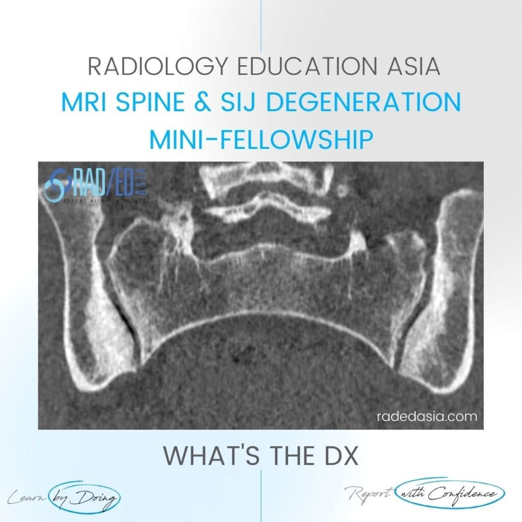
OSTEITIS CONDENSANS ILII RADIOLOGY CT SACROILIAC JOINT SIJ DISCUSSION ON OCI Osteitis Condensans Ilii. Typical CT sclerotic changes of Osteitis Condensans Ilii. The sclerosis is continuous with the SIJ margin and particularly on the patient’s right it has a triangular appearance. There is also mild sclerosis in the right sacrum adjacent to the […]
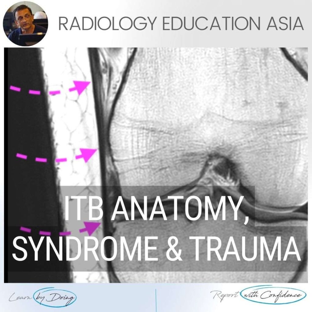
ILIOTIBIAL BAND MRI ANATOMY, SYNDROME, TRAUMA Finding and assessing the Iliotibial Band (ITB) on MRI of the Knee is the first structure we assess when looking at the lateral side of the Knee. In these three posts we look at: The normal MRI anatomy of the ITB. MRI of Iliotibial Band Syndrome (ITB Friction Syndrome)
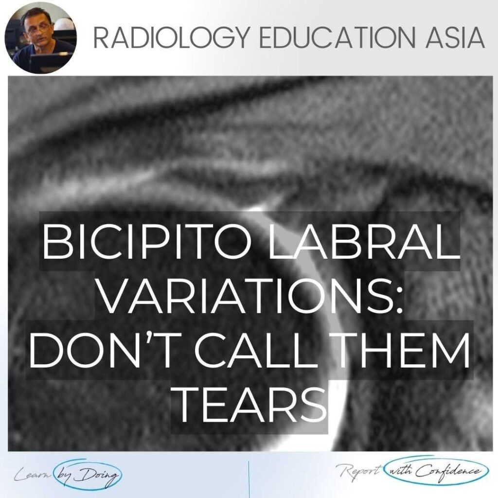
SHOULDER MRI LABRUM MRI VARIANT ANATOMY There are a number of normal appearances of the Bicipito Labral Complex which can mistakenly be called tears. Here is what they are and where to look for them. (These posts are Part of our Quick Pick Series with concise information from the Journals I read). CLICK
MRI DELAMINATING ROTATOR CUFF TEARS & MUSCULOTENDINOUS JUNCTION CYSTS On MRI Rotator cuff delaminating tears can result in Musculotendinous Junction Cysts. These Musculotendinous junction cysts are seen in the rotator cuff tendons, mostly in Supraspinatus and less commonly in infraspinatus. In this post we look at the appearance of both Delaminating Rotator cuff
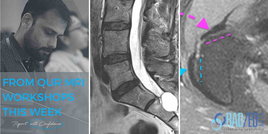
KEY POINTS FROM OUR SPINE MRI COURSES Spine MRI Online Radiology Course: Normal Spinal Enhancement Another three quick images with some basic key points from our online spine MRI courses. Here we look at areas of normal enhancement that can be mistaken for pathology: Normal enhancement of the anterior epidural plexus which can be mistaken
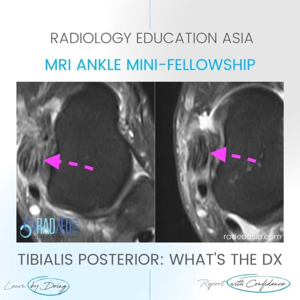
TIBIALIS POSTERIOR TENDINITIS TENDINOPATHY TEARS RADIOLOGY MRI WHAT ARE THE FINDINGS The tibialis posterior tendon is enlarged and increased in signal. DIAGNOSIS & DISCUSSION TIBIALIS POSTERIOR TENDINOPATHY AND PARTIAL TEAR. Tibialis posterior tendinopathy and delamination / partial tears. The normal tibialis posterior tendon should be uniformly low signal. Enlargement and or increased intermediate
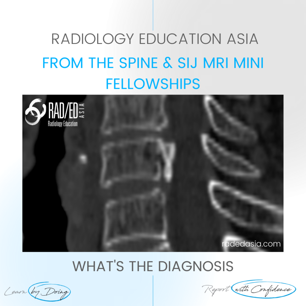
OPLL SPINE OSSIFICATION POSTERIOR LONGITUDINAL LIGAMENT CT DISCUSSION ON OPLL SPINE CT There is localised, linear ossification present at the posterior margin of the C6 vertebra. This has a typical appearance of OPLL (Ossification of the Posterior Longitudinal Ligament). The cervical spine is the most common location. It is important to follow
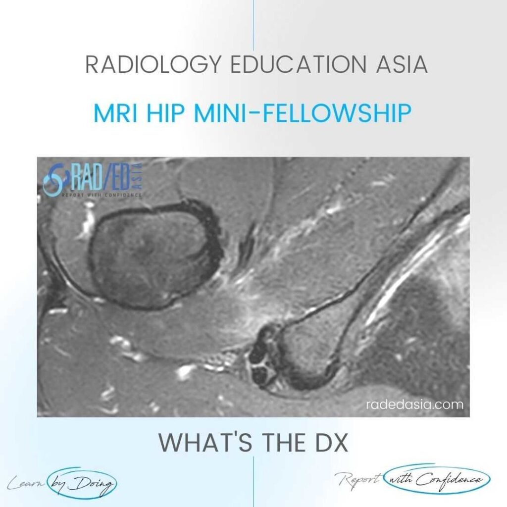
ISCHIOFEMORAL IMPINGEMENT HIP MRI RADIOLOGY ISCHIOFEMORAL IMPINGEMENT: WHY DOES IT OCCUR Ischiofemoral impingement is a result of the quadratus femoris muscle being compressed between the ischial tuberosity and the posterior femur resulting initially in quadratus femoris oedema but can progress to tears and severe atrophy. ISCHIOFEMORAL IMPINGEMENT: WHAT ARE THE FINDINGS Initially there is oedema
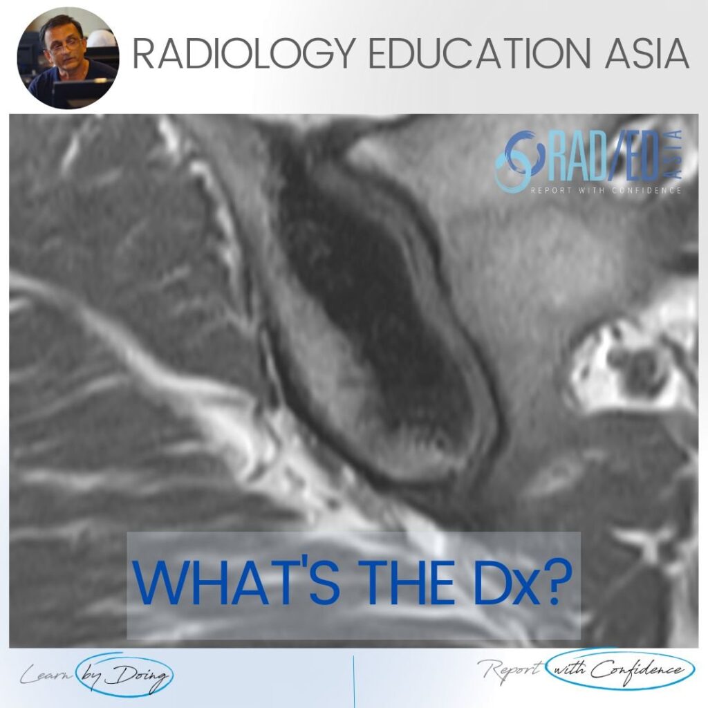
OSTEITIS CONDENSANS ILII MRI DIAGNOSIS OSTEITIS CONDENSANS ILII DISCUSSION OSTEITIS CONDENSANS ILII a common finding that may be confusing to diagnose on MRI. Look for low signal on all sequences The findings are predominantly of the iliac side of the SIJ but may be seen additionally on the sacral side. It doesn’t occur
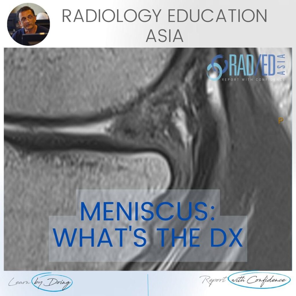
MENISCUS MACERATION DEGENERATION MRI KNEE WHAT ARE THE FINDINGS MENISCUS MACERATION DEGENERATION MRI KNEE Generalized increase in meniscal signal. Intermediate increase in signal on PD and PD/T2FS. The meniscus generally retains it’s shape but is ill defined. Due to the degeneration its more likely to tear and it also stops functioning like a normal
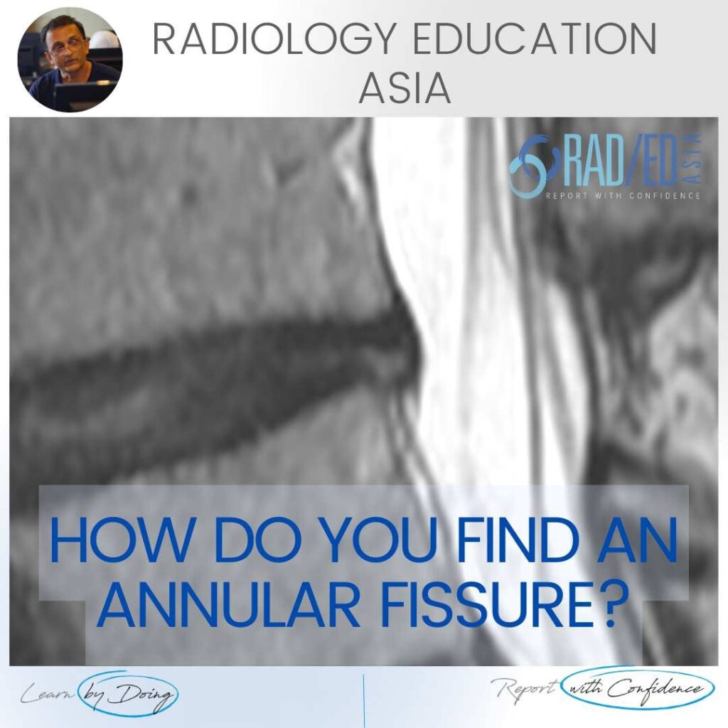
ANNULAR FISSURE TEAR MRI RADIOLOGY HOW DO YOU FIND AN ANNULAR FISSURE ANNULAR FISSURE ON MRI Annular fissures on MRI are relatively common. Whilst they don’t result in radiculopathy, they can be a cause of back pain and are important to diagnose and report. What does an annular fissure look like and where to
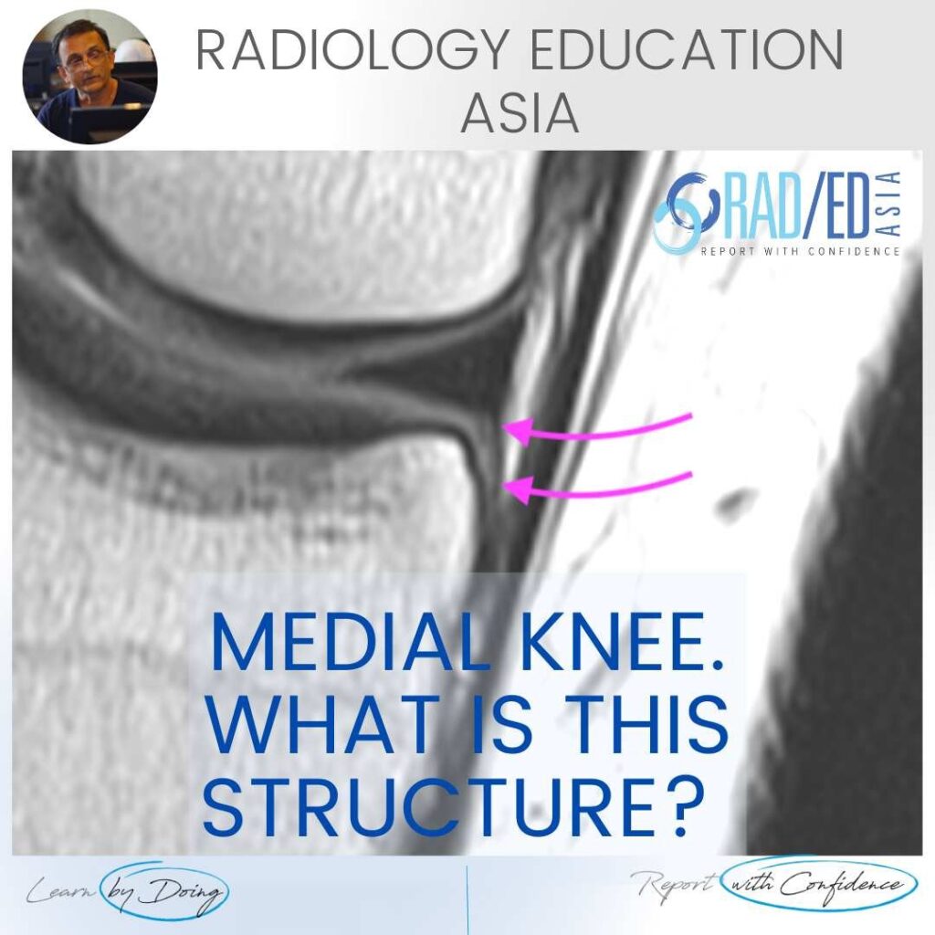
DEEP MCL ANATOMY KNEE MRI WHY BOTHER WITH RADIOLOGICAL ANATOMY? KNEE MRI MENISCO TIBIAL LIGAMENT DEEP MCL We stress understanding radiological anatomy in all our courses. Why? Because if you don’t know what normal looks like and where to look for it, you cant possibly work out what’s abnormal. See the video to know what
FATIGUE FRACTURE AND INSUFFICIENCY FRACTURE: WHAT'S THE DIFFERENCE DISCUSSION The difference lies in the underlying bone metabolism. Is it normal or not. A Fatigue fracture is stress type fracture due to repetitive overload, with normal bone metabolism (eg a March Fracture of the metatarsal in new army recruits). An Insufficiency fracture is a stress type
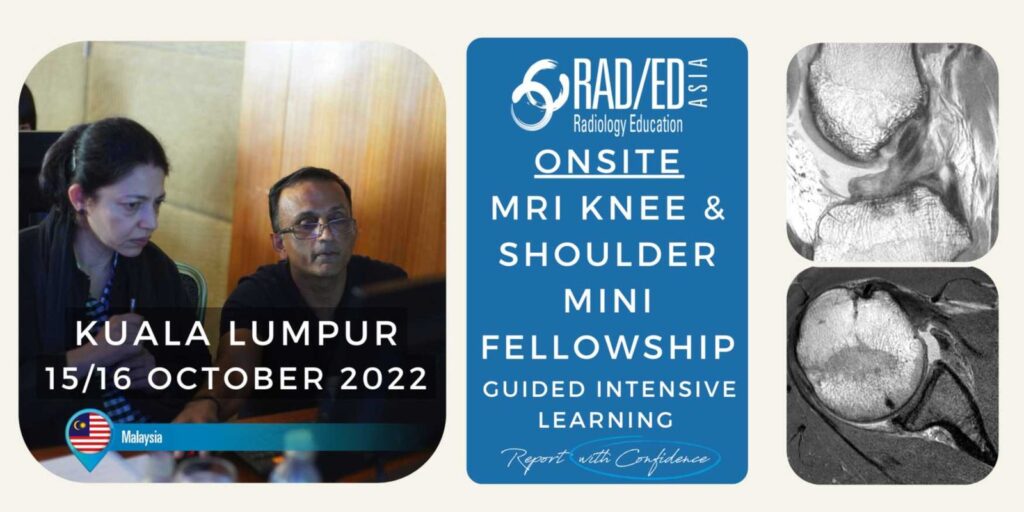
Home MSK RADIOLOGY CONFERENCE COURSE MUSCULOSKELETAL MRI MALAYSIA KUALA LUMPUR: MSK MRI RADIOLOGY KNEE & SHOULDER MINI FELLOWSHIP: 15/16 OCTOBER 2022 This Mini Fellowship is now over.To see our latest Onsite Guided Mini Fellowships please click HERE WHAT HAPPENED AT THE MINI-FELLOWSHIP @ KL CLICK ON THE IMAGE BELOW TO FIND OUT Our 2-Day Onsite
ANKLE IMPINGEMENT RADIOLOGY ANTERIOR X-RAY DISCUSSION Ankle impingement can occur at multiple sites. One of the more common ones is Anterior. The presence of Ankle impingement is ultimately a clinical diagnosis which is based on the patient’s symptoms and imaging findings, not just the imaging findings. So in the report we describe the
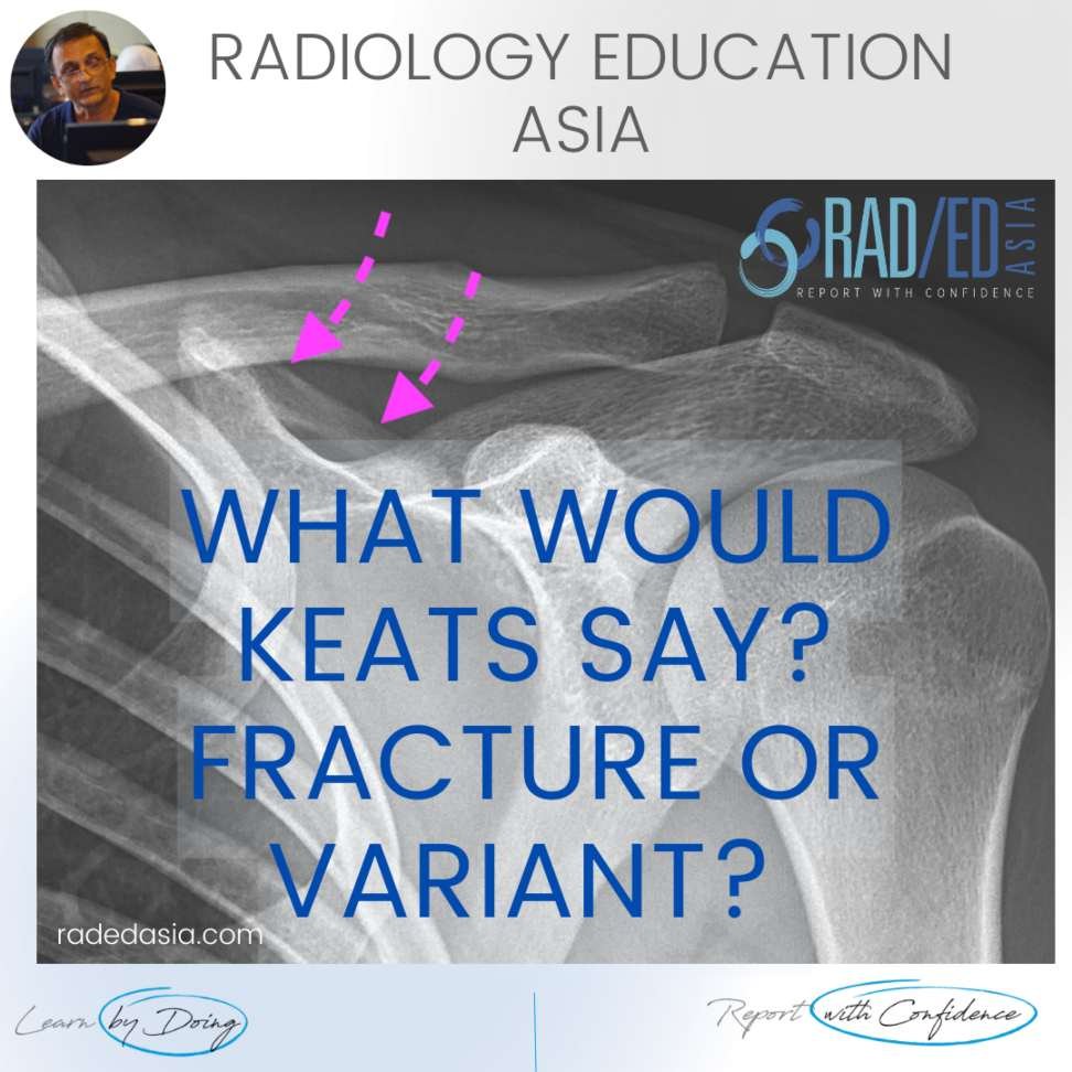
NORMAL SCAPULA VARIANT: CLASP LIKE SUPERIOR MARGIN FINDINGS Well corticated bridge of bone (Pink arrows) that lies on the superior margin of the scapula with a bone defect inferior to it. DISCUSSION This is a normal variant. Its described in Keats as a Clasp Like scapular margin that’s a result of bone being developmentally absent
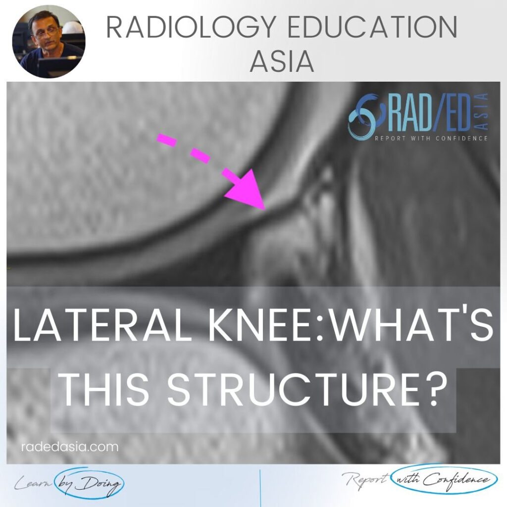
POPLITEOMENISCAL FASICLE MRI DISCUSSION: POPLITEOMENISCAL FASICLE MRI The popliteomeniscal fascicles are important structures to assess in knee trauma. There are three fasicles and the one shown here is the posterosuperior fascicle. It forms the roof of the popliteal hiatus. Tears of the fascicle can result in pain and lateral meniscal instability. https://youtu.be/pmWWMZWoctI If your Browser
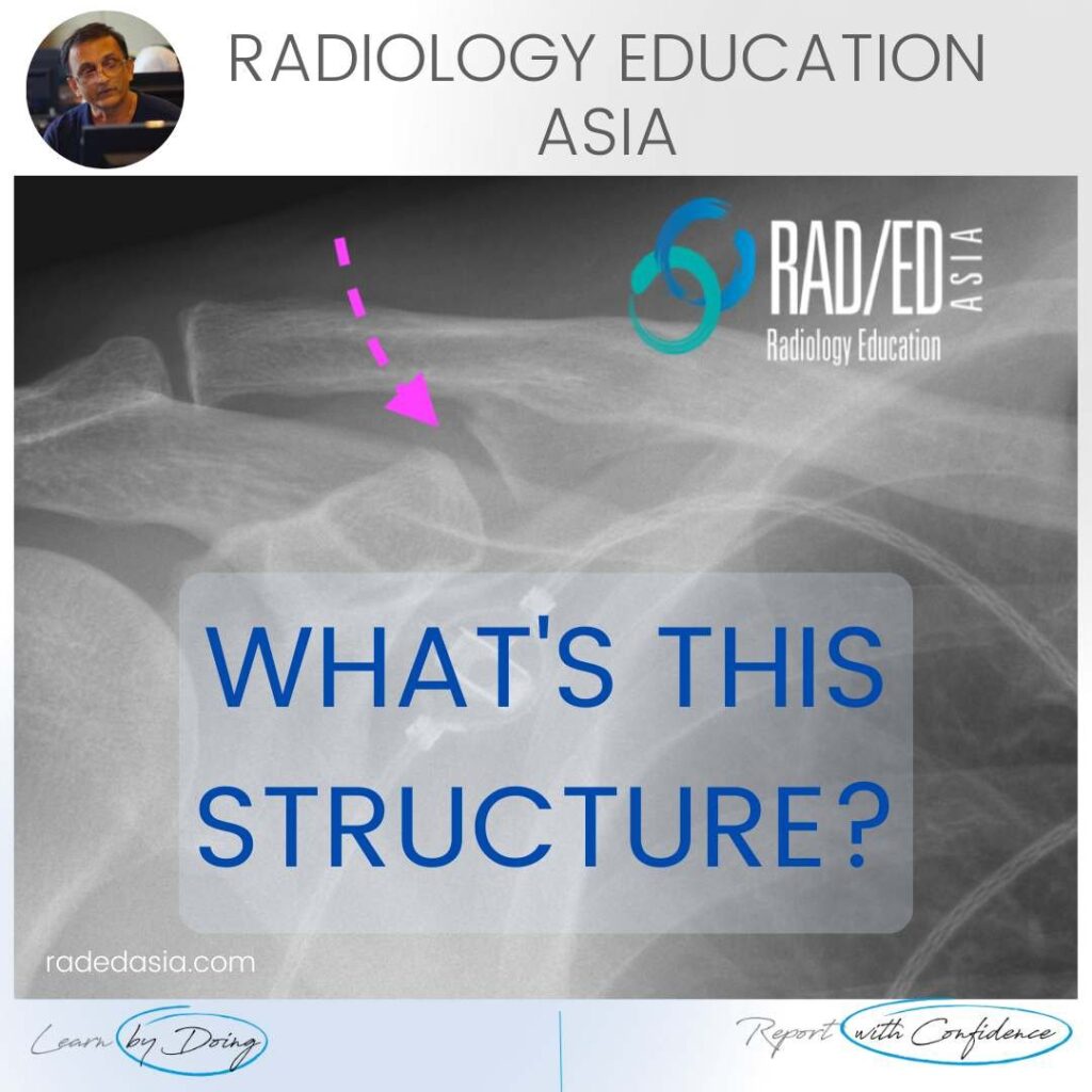
CORACOCLAVICULAR JOINT SHOULDER NORMAL VARIANT X-RAY CORACOCLAVICULAR JOINT NORMAL VARIANT ON X-RAY: The coracoclavicular joint is a normal variant where a synovial joint forms between the conoid tubercle of the clavicle and the coracoid process. The importance of recognizing this is that the joint can undergo degeneration which can be a source of
Home MSK RADIOLOGY CONFERENCE COURSE MUSCULOSKELETAL MRI INDIA BENGALURU: MSK MRI RADIOLOGY KNEE & SHOULDER MINI FELLOWSHIP: 17/18 SEPTEMBER 2022 THIS MINI-FELLOWSHIP IS NOW OVER CLICK HERE TO SEE OUR LATEST ONSITE MINI FELLOWSHIPS Our 2-Day Onsite Knee and Shoulder MRI Mini Fellowship and Workstation Workshop will be in Bengaluru, India on the 17th &
