CALCANEOFIBULAR LIGAMENT TEAR MRI ANKLE CALCANEOFIBULAR LIGAMENT NORMAL MRI The Calcaneofibular ligament is a small lateral ankle ligament that is often torn. Its normal MRI appearance is a thin low signal structure (Pink arrows) that extends from the calcaneum to the fibula and passes deep to the peroneal tendons (Green arrows). CALCANEOFIBULAR LIGAMENT […]
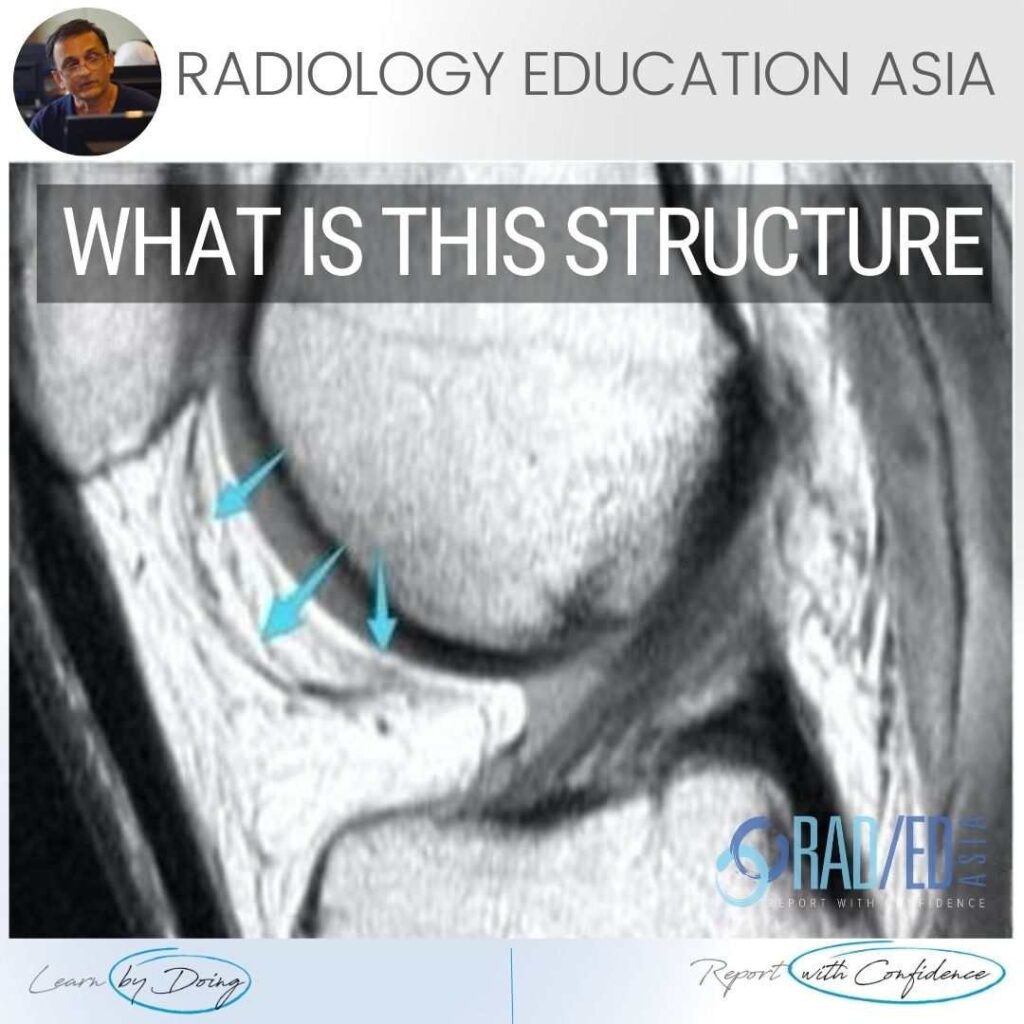
KNEE INFRAPATELLAR PLICA SYNDROME RADIOLOGY MRI DISCUSSION ON KNEE INFRAPATELLAR PLICA PLICAS IN THE KNEE The infrapatellar plica is one of 4 plicas in the knee: Medial Patella Plica. Suprapatellar Plica. Infrapatellar plica. Lateral Patellar plica. INFRAPATELLAR PLICA ANATOMY Attaches to anterior intercondylar notch and inferior pole patella. Runs through Hoffa’s Fat Pad.
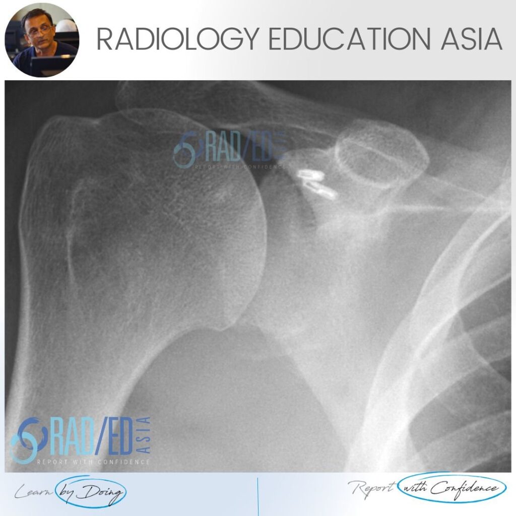
BONY BANKART LESION RADIOLOGY XRAY FINDING 1: BONE FRAGMENT Image Above: There is a thin fragment of bone at the inferior margin of the glenoid. FINDING 2: GLENOID FLATTENING Image Above: The inferior margin of the glenoid should normally be rounded and ovoid. Here it is flattened which is abnormal. DISCUSSION Bony Bankart lesions on
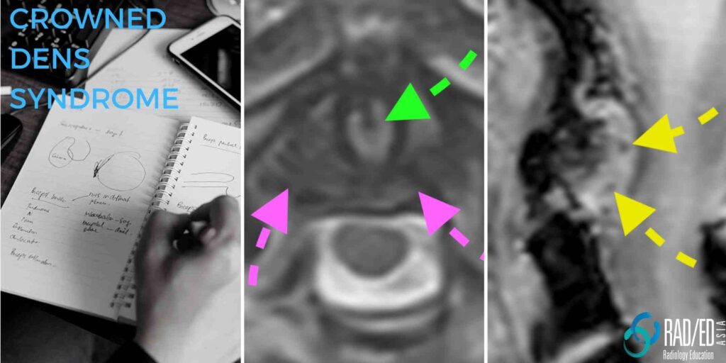
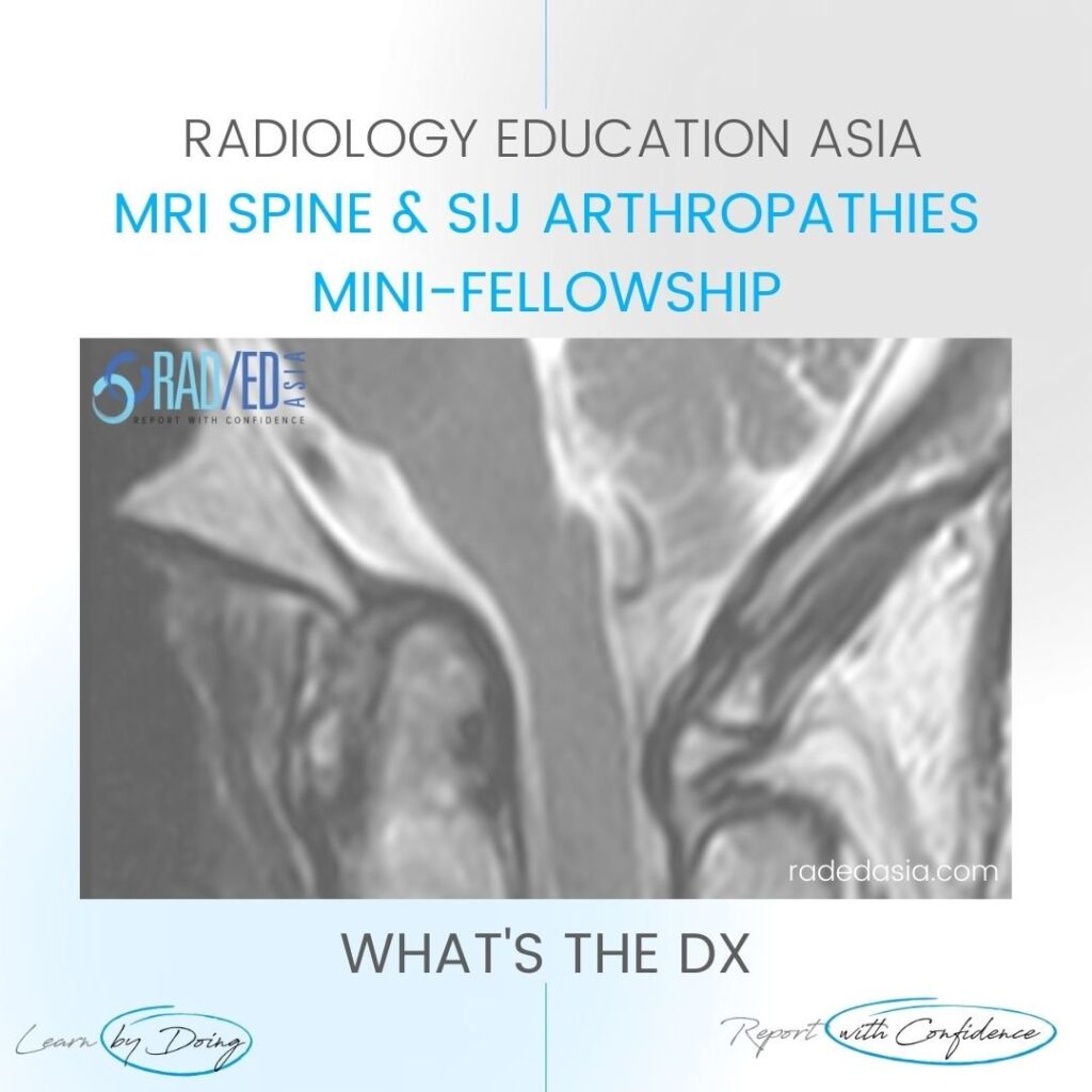
CPPD: CROWNED DENS SYNDROME CT AND MRI CPPD Crowned Dens Syndrome CT & MRI Spine Crowned dens is a manifestation of Calcium Pyrophosphate deposition diseases (CPPD) in the cervical spine. WHAT IS IT Peri odontoid deposition of Calcium Pyrophosphate crystals. WHAT DOES IT LOOK LIKE: CT The diagnosis is easier on CT as
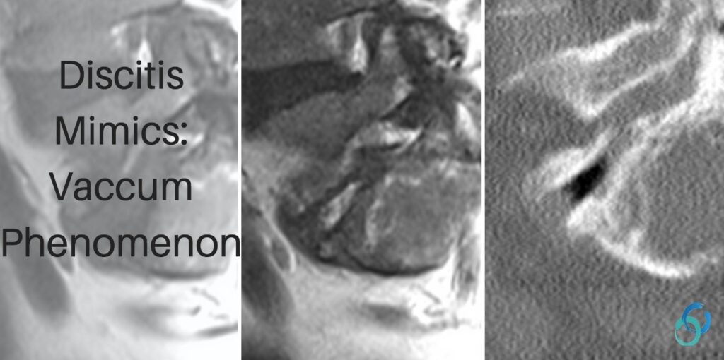
DISCITIS MIMICS: VACCUM PHENOMENON GAS IN THE DISC In discitis, one of the most important findings to look for is high T2 signal in the disc which alerts you to possible infection. But a degenerate disc with vacuum phenomenon (gas in the disc), can be high signal on T2 and mimic infection. Why does this
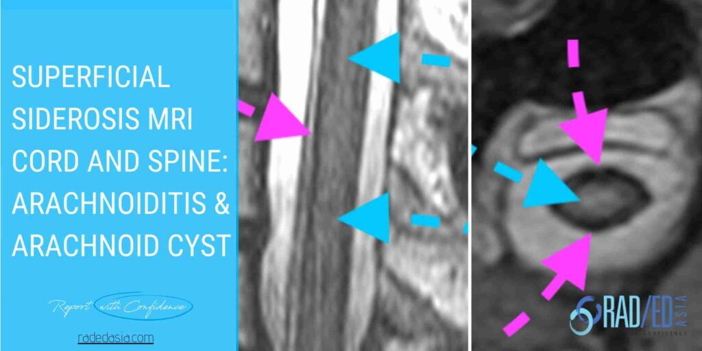
SUPERFICIAL SIDEROSIS: ARACHNOIDITIS & CYST Superficial Siderosis MRI Cord and Spine: Complications and Associations. Superficial Siderosis can affect the cord, nerve roots and the CSF space in the thecal sac. Here is what to look for and where to look to not miss this diagnosis. SUPERFICIAL SIDEROSIS MRI COMPLICATIONS TO LOOK FOR 1. ARACHNOIDITIS: WHY
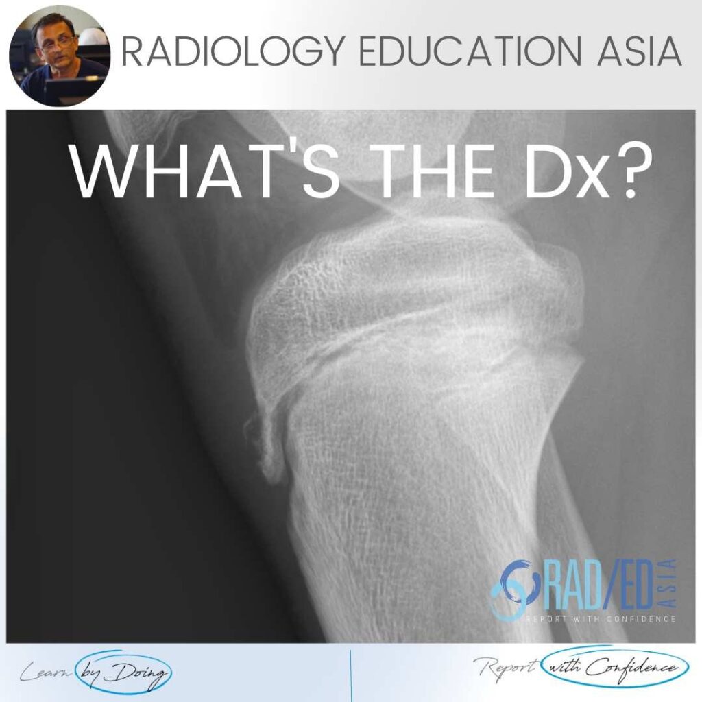
OSGOOD SCHLATTER RADIOLOGY X-RAY FINDINGS There are three findings in the x-ray that make the diagnosis ofOsgood Schlatter. Bone avulsion from tibial tuberosity. Soft tissue swelling over tibial tuberosity. Increased density deep infra patella bursa from fluid/inflammation. OSGOOD SCHLATTER X-RAY FINDINGS: VIEW VIDEO https://youtu.be/qG4b7ik9G3I If your Browser is blocking the video, Please view it on
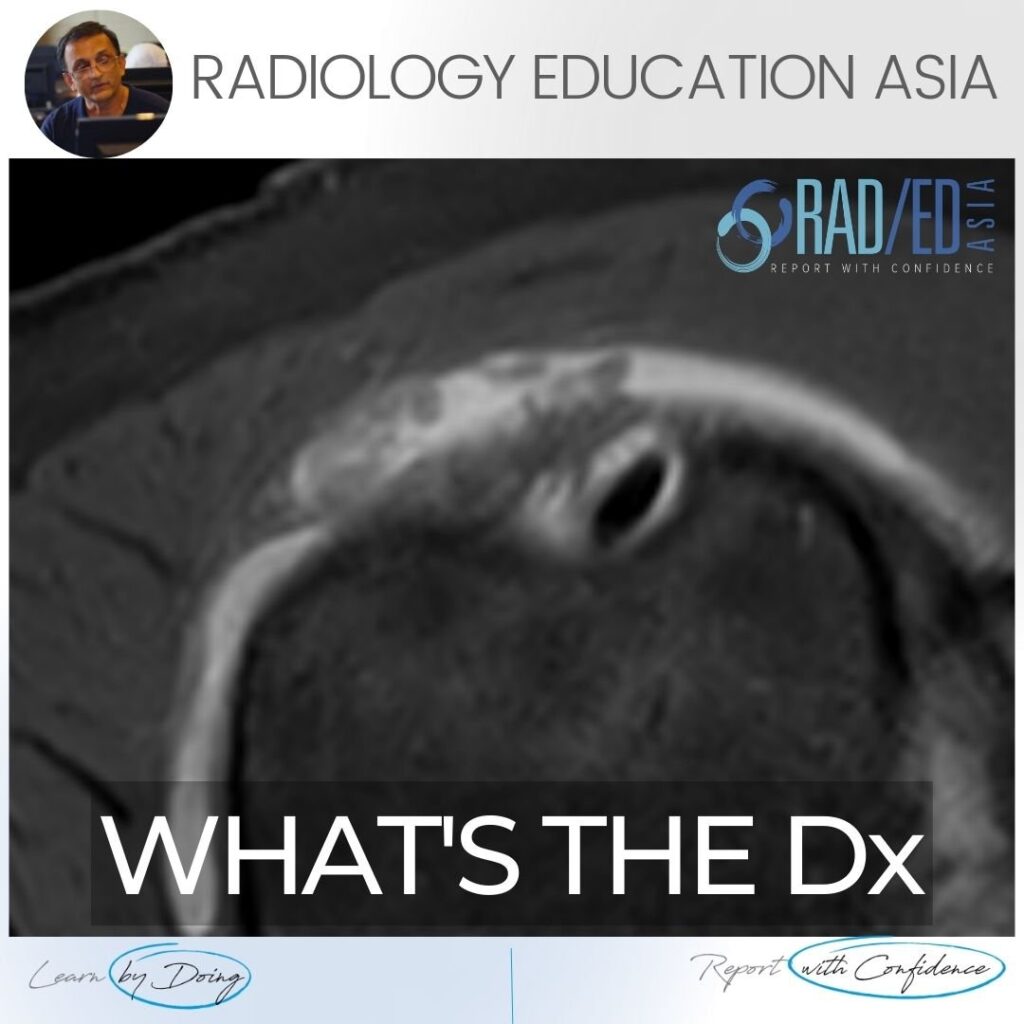
SUBACROMIAL SUBDELTOID BURSITIS MRI RADIOLOGY DISCUSSION Subacromial subdeltoid (SASD) bursitis on MRI can have a number of appearances. In this case we have a nodular type of synovitis which should raise the possibility of an inflammatory arthritis as a cause (Rheumatoid Arthritis in this case). WHAT DO WE SEE ON THE IMAGE? Fluid in the
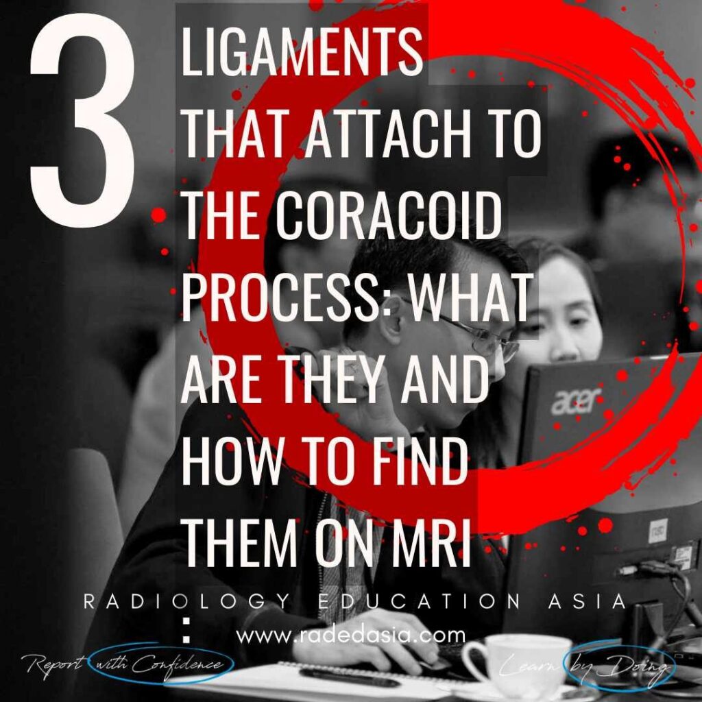
3 LIGAMENTS ATTACHED TO THE CORACOID PROCESS 3 LIGAMENTS ATTACHED TO THE CORACOID PROCESS: There are three ligaments that attach to the coracoid process of the shoulder. When reporting a shoulder MRI they should be part of the overall assessment. So what are they and how do you find them? See the brief video below.
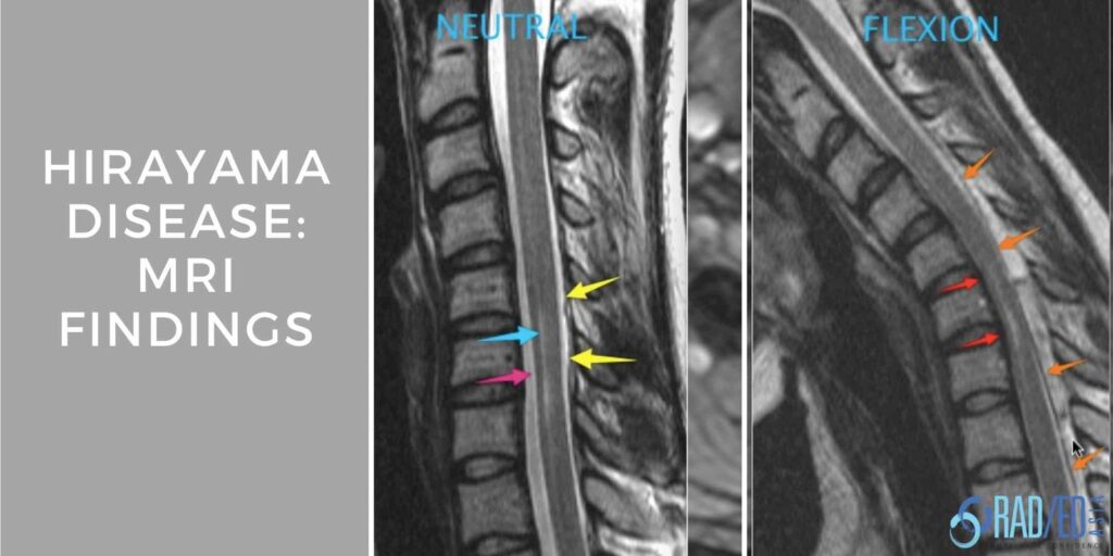
HIRAYAMA DISEASE MRI FINDINGS Hirayama Disease is a spinal muscular atrophy centered on the lower cervical and upper thoracic level which results in upper limb muscle wasting (amyotrophy). The presentation is chronic and occurs in adolescent young men with the condition being more common in Asia. The condition is caused by: Excessive anterior movement of
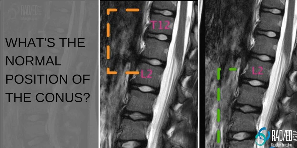
NORMAL CONUS POSITION THE NORMAL CONUS To determine if the cord is low lying or potentially tethered its important to know what is the lowest level you can see a conus and still call it normal. The radiology literature varies with a conus at the L2/3 disc or L3 level considered within the normal range.
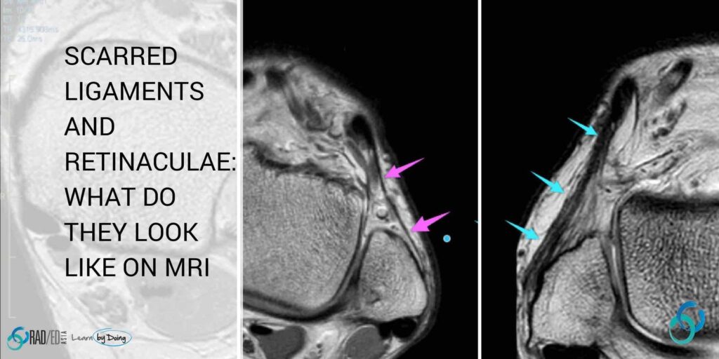
LIGAMENT AND RETINACULA HEALING: WHAT DOES SCAR TISSUE LOOK LIKE ON MRI NEED TO KNOW PATHOLOGY What happens to a ligament when it is torn and injured? This is a brief edited summary from an article on the pathology of scar formation ( “Scar formation and ligament healing” link below). “After an injury… hemostasis
Home MSK RADIOLOGY CONFERENCE COURSE MUSCULOSKELETAL MRI WORKSHOP UAE ABU DHABI: MSK MRI RADIOLOGY KNEE & SHOULDER MINI FELLOWSHIP: 25/26 FEBRUARY 2023 This Mini Fellowship is now over.To see our latest Onsite Guided Mini Fellowships please click HERE WHAT HAPPENED AT THE MINI-FELLOWSHIP @ ABU DHABI CLICK ON THE IMAGE BELOW TO FIND OUT Our
BASILAR INVAGINATION MRI RHEUMATOID ARTHRITIS BASILAR INVAGINATION MRI RHEUMATOID ARTHRITIS MCRAE'S LINE There can be apparent superior migration of the dens to the foramen magnum. It’s apparent because it’s due to cranial settling with the skull base moving down rather than the odontoid moving up. To assess this on imaging, there are a
Home MSK RADIOLOGY CONFERENCE COURSE MUSCULOSKELETAL MRI MALAYSIA KUALA LUMPUR: MSK MRI RADIOLOGY ANKLE, FOOT & HIP MINI FELLOWSHIP: 11/12 FEBRUARY 2023 This Mini Fellowship is now over.To see our latest Onsite Guided Mini Fellowships please click HERE WHAT HAPPENED AT THE MINI-FELLOWSHIP @ KL CLICK ON THE IMAGE BELOW TO FIND OUT Our 2-Day
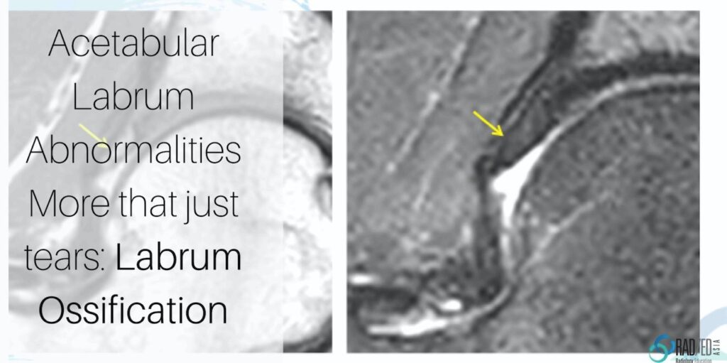
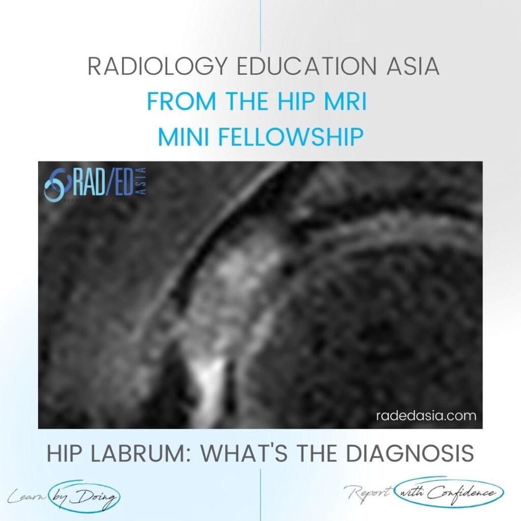
ACETABULAR HIP LABRUM OSSIFICATION: What does it look like Acetabular labrum ossification is not common and I have been able to find only a few reports on it. The cases we have seen mostly have an underlying dysplastic, elongated labrum however there are reports of it occurring in a normal labrum. So what does it
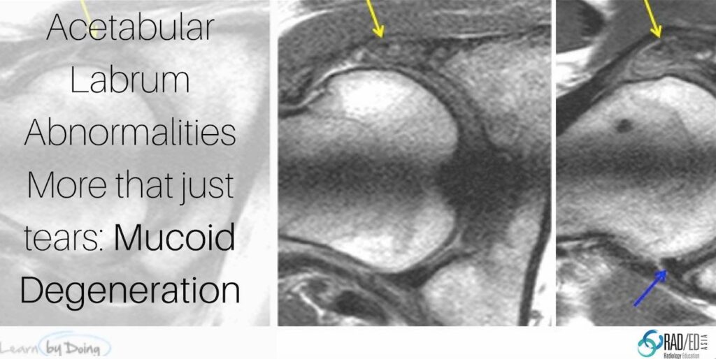
HIP ACETABULAR LABRUM MUCOID DEGENERATION There is a lot written about acetabular labral tears but there are other abnormalities of the labrum that can be precursors to or associated with tears. Here we look at Mucoid Degeneration. Mucoid degeneration of the acetabular labrum is not uncommon, but Ive not been able to find too much
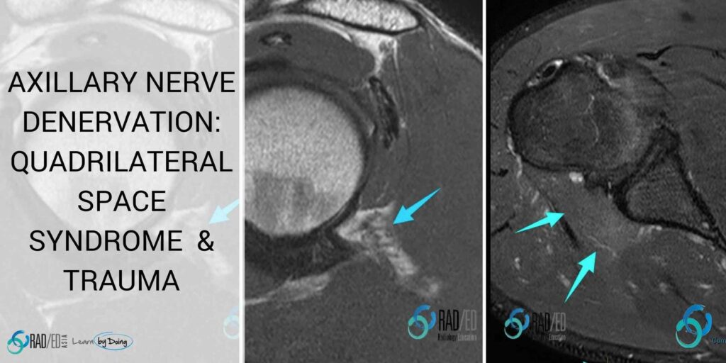
Denervation around the Shoulder: Quadrilateral Space Syndrome and Dislocation. This post looks at denervation of the Axillary Nerve in Quadrilateral Space Syndrome and Axillary nerve damage post shoulder dislocation both of which are less common causes of denervation around the shoulder. They have a particular distribution so recognizing the muscles involved gives you the
HIP LABRUM MUCOID DEGENERATION MUCOID DEGENERATION MRI APPEARANCE This is the MRI appearance of mucoid degeneration of the hip labrum. On MRI look for a number of findings Enlargement of the labrum Hyper-intensity of the labrum There may or may not be intra labral cysts of varying size. FINDINGS OF MUCOID DEGENERATION MRI Coronal PDFS
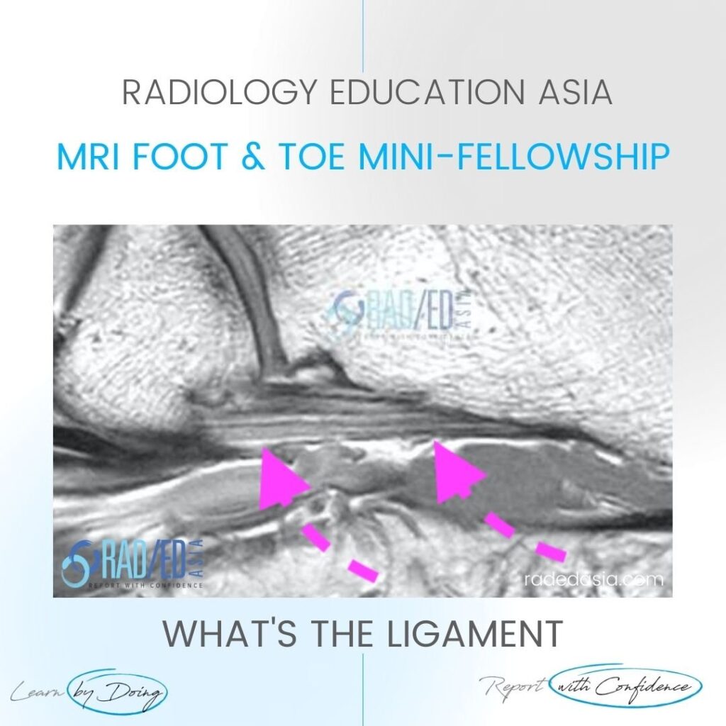
MRI PLANTAR LIGAMENT SHORT PLANTAR LIGAMENT WHAT'S THE STRUCTURE The Short Plantar Ligament. WHAT ARE THE FINDINGS ANATOMY OF THE SHORT PLANTAR LIGAMENT. 1. Extends from the calcaneus (Green star) to the cuboid (Yellow star). 2. It is distal and separate to long plantar ligament. 3. Has a fan shaped appearance. MRI PLANTAR LIGAMENT SHORT
SUBCORTICAL SUBCHONDRAL FRACTURE KNEE MRI TIBIAL PLATEAU DISCUSSION Subchondral or subcortical fractures of the knee can occur in the femoral condyles or tibial plateau. The key to the diagnosis is seeing a linear low signal line adjacent to and paralleling the cortex without any cortical defect or break in the acute phase. There
