Antero Medial Ankle Impingement MRI
| Pathology |
|
The causes of antero medial impingement are the same as those we have discussed in the previous posts of other impingement locations.
- Granulation tissue, synovitis and scar formation from trauma to the anterior fibres of the deep deltoid ( Anterior Tibio Talar Ligament)
- Anteromedial Capsule tear and scar
- Spur formation

|
Image Above: Pink arrows anterior fibres deep deltoid , blue arrow posterior fibres deep deltoid. Note that the anterior fibres are much thinner and at time may not be seen on MRI.
MRI Appearance
|
|
 Image Above: Hypertrophic scar formation deep and superficial fibres of the deltoid ( blue arrows) extending between Tibialis Posterior ( red arrow) and FDL tendon ( yellow arrow). This is a severe case of scarring. Image Above: Hypertrophic scar formation deep and superficial fibres of the deltoid ( blue arrows) extending between Tibialis Posterior ( red arrow) and FDL tendon ( yellow arrow). This is a severe case of scarring.
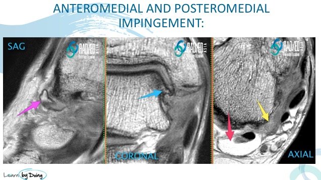
 Image Above: Spur formation from the medial margin of the talus ( pink and blue arrows) with joint space narrowing on the coronal scan. Important to look at the extreme sagittal scans covering the anteromedial region as the spurs are often better seen on the sagittal scans. Also granulation tissue in the posteromedial gutter ( yellow arrow) and FHL tenosynovitis ( red arrow). Image Above: Spur formation from the medial margin of the talus ( pink and blue arrows) with joint space narrowing on the coronal scan. Important to look at the extreme sagittal scans covering the anteromedial region as the spurs are often better seen on the sagittal scans. Also granulation tissue in the posteromedial gutter ( yellow arrow) and FHL tenosynovitis ( red arrow).
|


