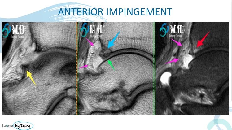
ANTERIOR ANKLE IMPINGEMENT MRI
| Need to Know: Anatomy and Pathology |
|
Anterior Ankle Impingement is caused by repeated ankle dorsiflexion with resulting micro trauma to the anterior tibial plafond and corresponding talar neck. This results in bone damage and periosteal bleeding leading to proliferative bone changes and spur formation. The bony spurs are not due to traction osteophytes at the attachment of the dorsal capsule. Secondary anterior capsular thickening and scarring, synovitis and loss of anterior joint cartilage develop. |
| What to look for. |
|
1.Bone spur anterior tibial plafond and talar neck 2.Anterior capsular thickening 3.Anterior Synovitis 4.Cartilage loss anterior margin 5.Bone marrow oedema in spurs Image Above: All the changes seen in anterior ankle impingement. Talar neck spur ( yellow arrow), Anterior Tibial Plafond Spur ( blue arrow), Capsular Thickening and Synovitis ( Pink Arrows), Bone Marrow Oedema at spur ( red arrow) and Chondral loss ( green arrow). |


