ACROMIAL APOPHYSIOLYSIS OR APOPHYSITIS MRI FINDINGS
WHAT IS ACROMIAL APOPHYSIOLYSIS
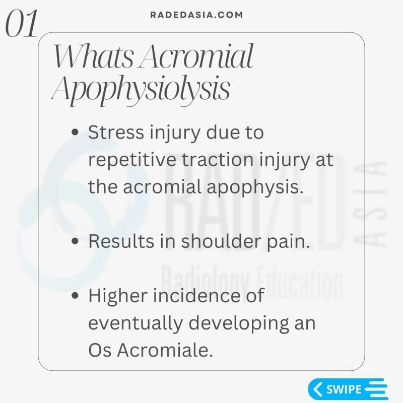
- Acromial apophysiolysis or acromial apophysitis is a Stress injury due to repetitive traction injury at the acromial apophysis.
- It results in instability at the apophysis and shoulder pain.
- People who have acromial apophysitis have a higher incidence of eventually developing an Os Acromiale.

- The acromion develops from 3 apophyses which progressively fuse by age 25.
- <25yo unfused apophysis is normal but there should be no bone marrow oedema around the apophysis.
- If >25yo and apophysis is unfused unfused = Os Acromiale. (Discussed this and how to find it in a previous post you can see HERE

3 MRI FINDINGS IN ACROMIAL APOPHYSIOLYSIS
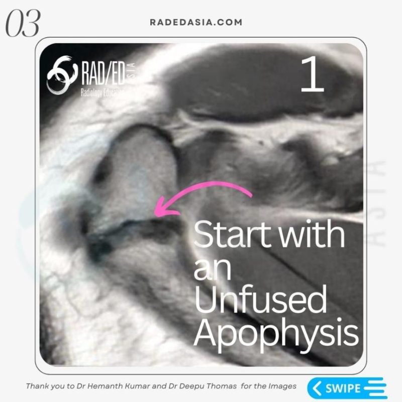
- The apophysis has to be unfused.
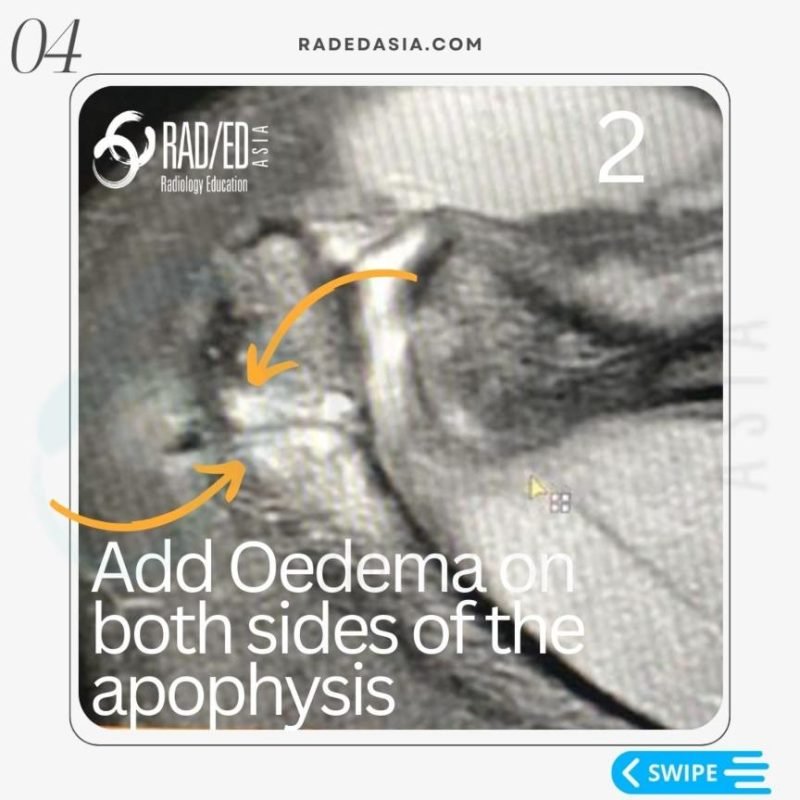
- Oedema on both sides of the apophysis.
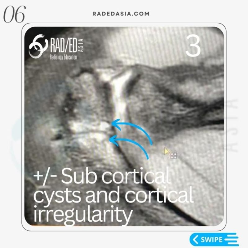
- +/- Cystic type change and cortical irregularity subjacent to growth plate.

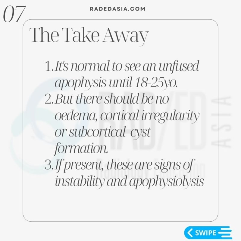
- It’s normal to see an unfused apophysis until 18-25yo.
- But there should be no oedema, cortical irregularity or subcortical cyst formation.
- If present, these are signs of instability and apophysiolysis.
See the images below for what the MRI appearance of Acromial Apophysiolisis looks like.

ACROMIAL APOPHYSIOLYSIS OR APOPHYSITIS: VIEW IMAGES


Previous
Next
Learn more about SHOULDER Imaging in our ONLINE
Guided MRI SHOULDER Mini-Fellowship.
More by clicking on the images below.
- Join our WhatsApp Group for regular educational posts. Message “JOIN GROUP” to +6594882623 (your name and number remain private and cannot be seen by others).
- Get our weekly email with all our educational posts: https://bit.ly/whathappendthisweek
OTHER POPULAR POSTS ON SHOULDER MRI: CLICK ON THE IMAGE BELOW
#radedasia
#radiology #radedasia #mri #shouldermri #msk #mskmri #radiologyeducation #radiologycases #radiologist #rads #radiologystudent #radiologycme #radiologycpd #medicalimaging #imaging #radcme #rheumatology #arthritis #rheumatologist #sportsmed #sportsphysician #orthopaedic #osacromiale #apophysiolysis #acromialapophysitis

