MRI Features of Spine Pathological Fractures
In this post we look at the MRI findings that suggest a Pathological Fracture.
| FINDINGS THAT SUGGEST A PATHOLOGICAL FRACTURE |
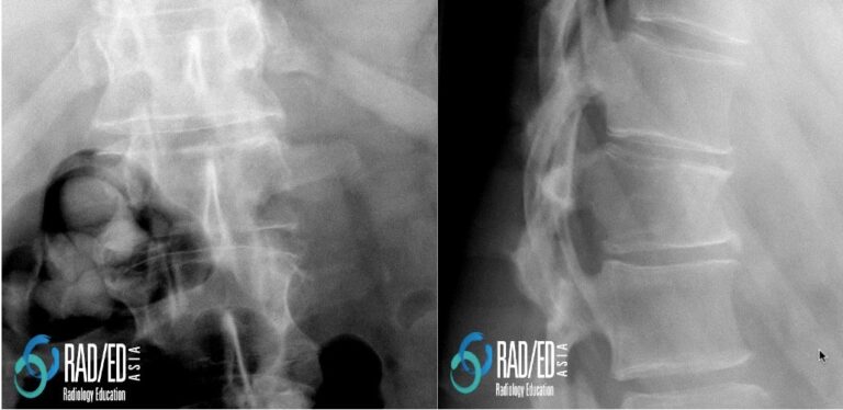 Image Above: L1 compression fracture. Benign or malignant. See MRI images below on how to differentiate. Image Above: L1 compression fracture. Benign or malignant. See MRI images below on how to differentiate.
WHAT TO LOOK FOR ON MRI
- Convex Posterior Margin
- Abnormal marrow signal in the pedicle
- Soft tissue in the anterior epidural space
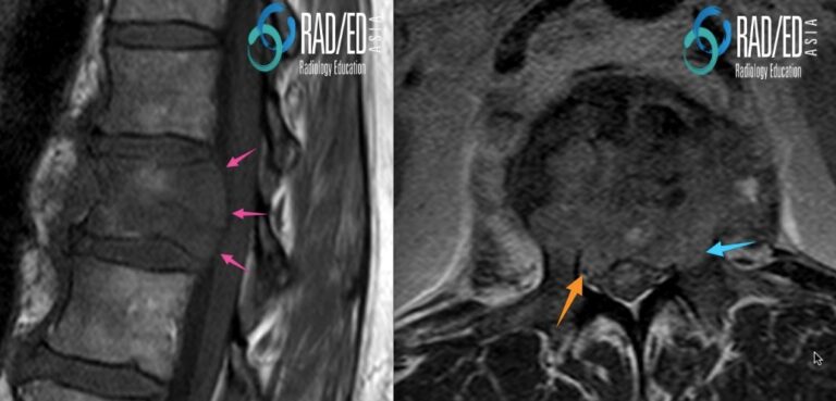
Image Above: Pathological Fracture demonstrating a convex posterior margin ( pink arrows), extension into the left pedicle ( blue arrow) and anterior epidural extension ( orange arrow). Almost complete reduction in T1 signal of the vertebral body is also suggestive of infiltration but less convincing than the findings above.
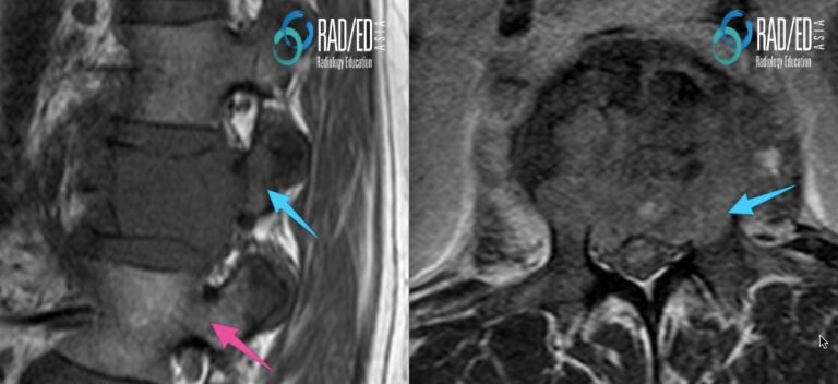 Image Above: How to assess the pedicles on sagittal scans. Look on the sagittal images for the marrow signal in the pedicles in addition to the axial images. Pedicle involvement ( blue arrows) compared to a normal pedicle ( pink arrow). Image Above: How to assess the pedicles on sagittal scans. Look on the sagittal images for the marrow signal in the pedicles in addition to the axial images. Pedicle involvement ( blue arrows) compared to a normal pedicle ( pink arrow).
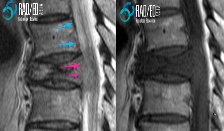
Image Above: Loss of posterior cortex black signal ( pink arrows). Compare with normal posterior cortical margin ( blue arrow). |
IS IT FOOL PROOF?
|
| No, as not all pathological fractures will have these findings but if they are present it makes it strongly suggestive.
You need to look at all the findings and then make a decision as to whether it falls more into the benign or pathological camp.
|
WHAT TO DO IF IT’S NOT CLEAR?
|
|
At times it will not be possible to make a definite decision as the findings are indeterminate if its benign or malignant. What to do then
- Check the remainder of the spine for further lesions.
- Bone scan to look for any other sites
- Chest Xray
If all of those end up negative then the two options remaining are for a biopsy or wait and rescan to see if it evolves like a benign or pathological fracture.
|

