MRI Wrist and Finger: Sagittal Band
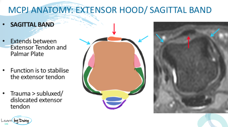
The sagittal band is part of the extensor hood of the fingers and serves to hold the extensor tendon of the finger in place. In this post a quick review of the anatomy and the appearance of a tear of the sagittal band. The anatomy is complicated so the aim of this post is for you to have a rough understanding of the appearance of the Sagittal band on these static images. In the workshop we will go though how to identify it on the dicoms.
KEY POINTS:
- Is part of the extensor hood but surrounds the MCPJ.
- It surrounds the extensor tendon on the dorsal aspect of the joint and on the volar side is continuous with the Volar Plate.
- On MRI the normal appearance is of a thin black line surrounding the joint.
Image Above: Sagittal band ( blue arrows) extends between the extensor digitorum tendon ( red arrow). It attaches to the Volar Plate ( yellow on the diagram). Low signal on all sequences and is thin and well defined.
Image Above: Normal sagittal band ( blue arrows).
Image Above: Torn sagittal band ( blue arrows) ill defined thickened and hyperintense. Normal sagittal band ( red arrows).


