MRI Finger Collateral Ligaments
MRI Finger Collateral ligaments. Quick review of the appearance of the ulnar and radial collateral ligaments of the fingers. The examples are of the MCPJs but the same appearance applies for the PIP and DIPJs. We look at normal, tears, avulsions and periosteal stripping.
ANATOMY:
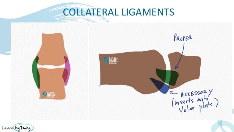
- There is a radial and ulnar collateral ligament on either side of the MCPJ.
- Each has a Proper and an Accessory component.
- The Proper portion attaches to bone on either end.
- The Accessory portion attaches to the Metacarpal proximally and to the Palmar/ Volar Plate distally.
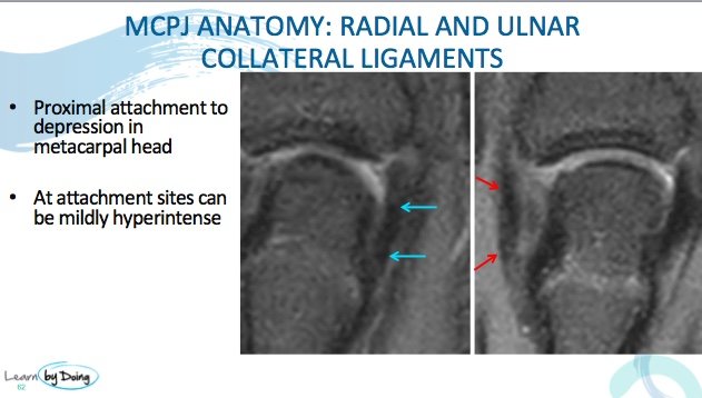
Image Above: Normal appearance of the UCL and RCL. Low signal on all sequences and taut like all ligaments.
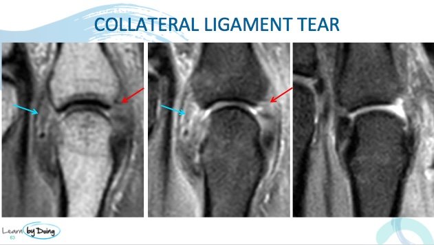
Image Above: Tears of the UCL and RCL demonstrating thickening and hyperintensity in keeping with tears. No retraction to suggest avulsion.
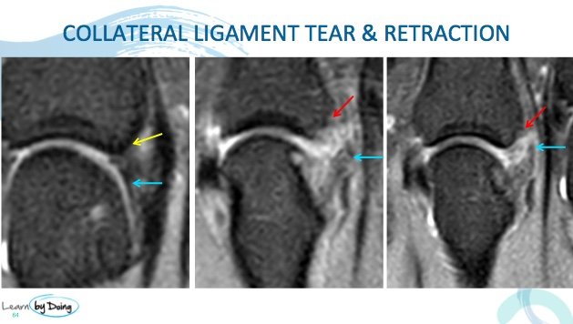
Image Above: First image demonstrates normal RCL and attachment. Tear of the RCL demonstrating thickening and hyperintensity and retraction ( blue arrow) and site of avulsion ( red arrow).
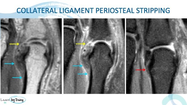
Image Above: Tear of the UCL demonstrating thickening and hyperintensity and avulsion ( yellow arrow) and stripping of the proximal attachment with hemorrhage underneath ( blue arrow). Last image demonstrates normal UCL attachment.

