msk mri radiology conference

MRI Wrist Scaphoid Fractures

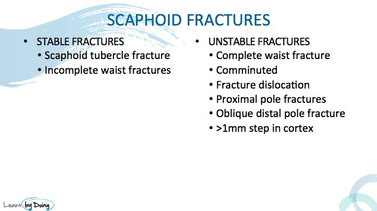
The various appearances of scaphoid fractures and contusions on MRI and an understanding of why fractures in certain areas are more likely to undergo non union or AVN because of theĀ  the vascular supply of the scaphoid.

mri wrist scaphoid fracture radiology education asia

mri wrist scaphoid contusion radiology education asia

Image Above: Scaphoid contusion ( blue arrow) is diagnosed by seeing oedema from bone bruising but no fracture line.

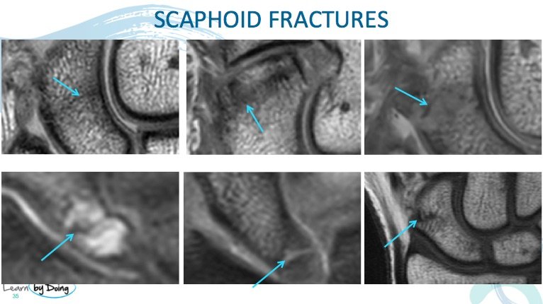
mri wrist scaphoid fractures radiology education asia.png

Image above: Various appearances of scaphoid fractures.


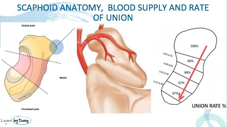
mri wrist scaphoid fracture vasuclar supply radiology education asia

Image above: Vascular supply to the scaphoid is retrograde so the more proximal a fracture is, the higher the chance of non union developing or AVN. Complications more likely with proximal pole fractures and less likely with distal pole fractures. ( Images unknown source).

Leave a Comment

Radiology Education

Join our Newsletter

Stay tuned on new
Mini-Fellowships launches and learnings

Mini-Fellowships

Better Than Conferences

We help radiologists learn by doing and report with confidence

Scroll to Top

Log in to your Mini-Fellowship or create an account

Mini-Fellowships

Better Than Conferences

We help radiologists
learn by doing and
report with confidence

Join our Newsletter

Stay tuned on new
Mini-Fellowships launches and learnings

Mini-Fellowships

Better Than Conferences

Join our WhatsApp Community

We help radiologists learn
by doing and report with confidence