MRI Hip: Bursae around the Hip
Although we mostly talk about the trochanteric bursa, there are a number of bursa around the trochanter and hip which can get inflamed. This is a review of the bursa that can be seen on Hip MRI.
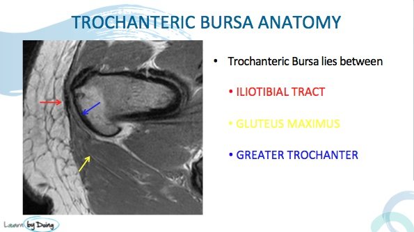
- Trochanteric Bursa
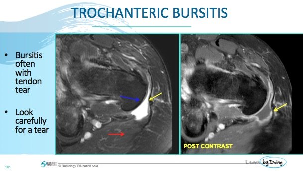
Image Above: Trochanteric bursitis ( yellow arrow). Bursa lies in-between greater trochanter ( blue arrow), Gluteus Maximus ( red arrow) and the Iliotibial Tract ( black structure at tip of yellow arrow). Post contrast there is enhancement of the margins of the bursa in keeping with inflammatory change.
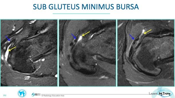
2. Sub Gluteus Minimus Bursa
Image Above: Sub Gluteus Minimus bursa ( yellow arrow) deep to gluteus minimum tendon ( blue arrow).
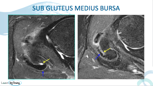
3. Sub Gluteus Medius Bursa.
Image Above: Sub Gluteus Medius bursa ( yellow arrow) deep to gluteus medius tendon ( blue arrow).
4. Iliopsoas Bursa.
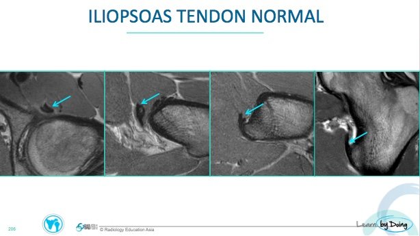
Image Above: Normal ilio psoas tendon ( blue arrow). Normally will not see any fluid around the tendon.
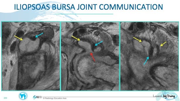
Image Above: Iliopsoas tendon ( blue arrow) surrounded by the iliopsoas bursa ( yellow arrow). Note normal communication between joint and bursa ( red arrow). If there is a joint effusion it can decompress into teh iliopsoas bursa.
5. Obturator Internus Bursa
Image Above: Fluid seen in Obturator interns bursa ( blue arrow) lying between the ischium and obturator interns ( yellow arrows).

