Hip MRI Gluteal Tendon Abnormalities
MRI Hip Gluteal tendons. This post reviews the various abnormalities seen of the gluteal tendons from tendinosis and tears to the feature of avulsion.
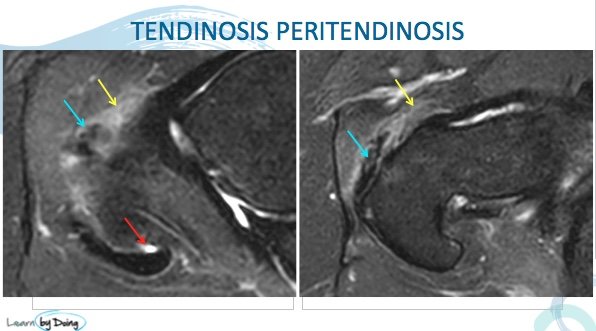
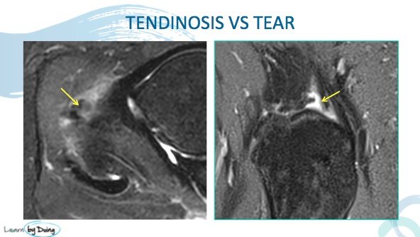
Image Above: Gluteus minimus tendinosis ( blue arrow) with ill definition and mild increased signal of the tendon. Peri tendinosis which is inflammatory change around the tendon ( yellow arrow). Small amount of fluid in th sub gluteus medium bursa ( red arrow).
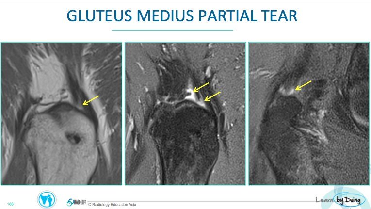
Image Above: Partial tear posterior fibres of gluteus medius ( yellow arrow). Interruption of black tendon fibres with fluid signal indication fluid accumulating in a tear.
3 Features of a Complete Tear and Avulsion:
- Bare Facets
- Tendon Laxity
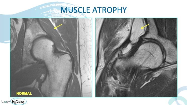
- Muscle atrophy
Image Above: Complete tear and avulsion resulting in bare facets ( no tendon attachment seen) of gluteus minimum ( yellow arrow), anterior fibres of gluteus medium ( red arrow). Posterior fibres of gluteus medium ( blue arrow) still attached but demonstrate tendinosis with mild increased signal.
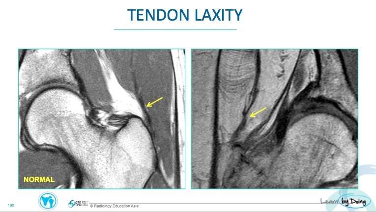
Image Above: Image on right demonstrates laxity of the anterior gluteus medium tendon ( yellow arrow) which indicates that there has been rupture of the fibres from their attachment. Compare with normal taut tendon on left.
Image Above: Atrophy of the gluteus minimum muscle ( image on right yellow arrow) compare with normal on left image. Significant atrophy like this usually indicates underlying rupture of the tendon.

