MRI Knee Meniscal Degeneration : What differentiates it from a meniscal tear
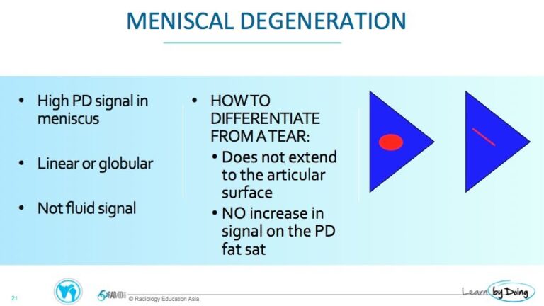
Meniscus degeneration on MRI can sometimes be difficult to differentiate from a meniscal tear. Review of the features of meniscal degeneration and how to distinguish them from tears.
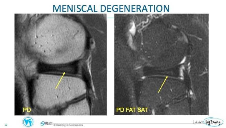
Image Above: Linear high signal ( yellow arrow) that does not extend to an articular surface = degeneration.
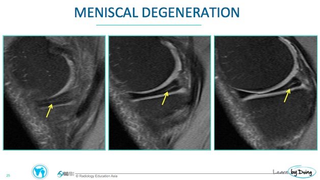
Image Above: Globular high signal ( yellow arrow) that does not extend to an articular surface = Degeneration.
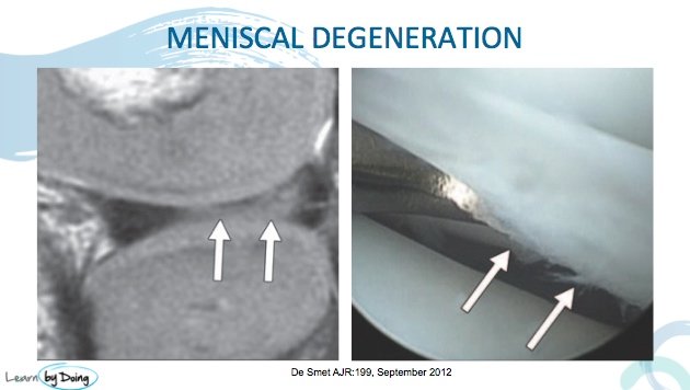
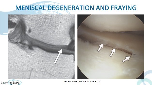
Two Images above: I found these slides really useful in visualising what is actually seen on arthroscopy of what we report as degeneration on MRI. The surface on the arthroscopy image is frayed but no tear. On MRI we don’t see the fraying because of lack of resolution. All images from De Smet et al AJR 199 September 2012.
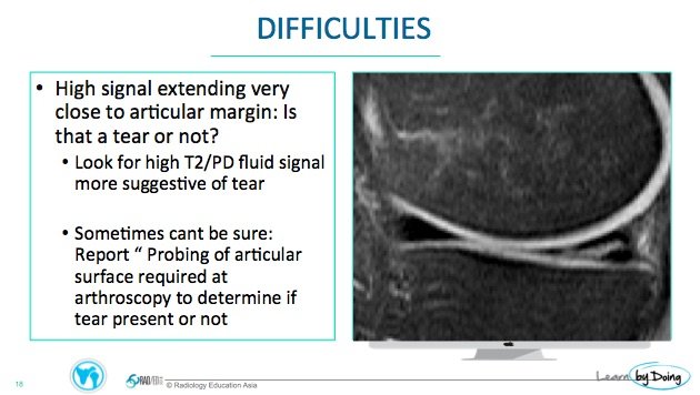
Sometimes though its not as straight forward as above. It can be difficult to be certain if it is all degeneration or there may be a small tear.

