Shoulder MRI : Rotator Cuff Tendon Delamination
There are three appearances that can be seen on MRI when there is delamination of the rotator cuff tendons. If we keep in mind that delamination is essentially a longitudinal separation and peeling of tendon fibres with the area of separation accumulating fluid.
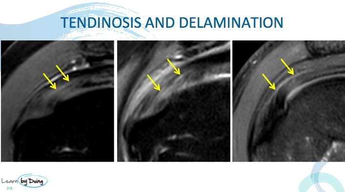
- In moderate to severe tendinosis there can be thin, longitudinal separation of tendon fibres that is not considered a tear.
Image Above: Diffuse increase in supraspinatus tendon in keeping with tendinosis. Areas of linear more fluid type signal ( yellow arrows) are areas of delimitation or longitudinal separation of tendon fibres.
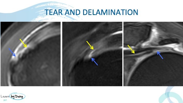
2. Intrasubstance delaminating extension of a burial or articular surface tear.
Image Above: Tears of SST ( blue arrows) which have propagated as delaminating tears ( yellow arrows) into the substance of the tendon.
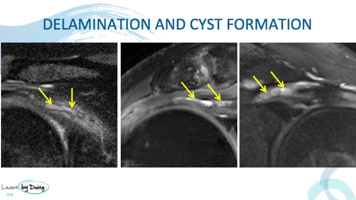
3. Intramuscular/ musculotendinous junction cyst formation. At the musculotendinous junction the delamination is arrested and fluid accumulates and cyst formation begins.
Image Above: Delaminating tears as they get to the musculotendinous junction form cysts ( yellow arrows) . Any cyst at the musculo tendinous junction should alert you to the presence of a delaminating tear.


