SLAP 4 Tears The best way to find biceps extension of the tear on MRI
In the workshop looking at SLAP tears we stressed finding and describing the abnormalities on MRI rather than memorizing the infinite numbers of SLAP lesions. So when a SLAP tear extends from the labrum into the biceps tendon the best way to assess this is on the sagittal PD scans. This is because as we discussed, the intra articular portion of the biceps tendon is best seen on sagittal scans because the tendon runs perpendicular to the scan plane. Look for high signal, that approximates fluid signal, in the biceps tendon at and adjacent to its insertion.
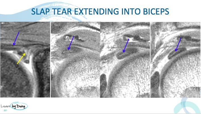
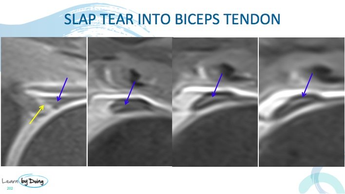
Image Above: SLAP tear ( yellow arrow) extending into the biceps tendon ( blue arrows). First image coronal remainder are sagittal through the biceps tendon adjacent to its insertion. Coronal image high signal tear ( blue arrow) seen in biceps tendon but because of partial voluming it can often be difficult to be certain. The sagittal scans show definite increased signal within the tendon in keeping with extension of the labral tear into the biceps tendon.
Image Above: SLAP tear ( yellow arrow) extending into the biceps tendon ( blue arrows). First image coronal remainder are sagittal through the biceps tendon adjacent to its insertion. Coronal image high signal tear ( blue arrow) seen in biceps tendon but because of partial voluming it can often be difficult to be certain. The sagittal scans show definite increased signal within the tendon in keeping with extension of the labral tear into the biceps tendon.


