MRI Subscapularis Tears
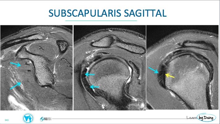
MRI of Subscapularis tears can be confusing to assess only on the axials. We review the sagittal location to look for tears and what they look like. Remember, look at the sagittals first for where the subscapularis inserts ( humeral head surface becomes flat and angled) and is 1 slice medial to the bicipital groove.
Image above: Normal Subscapularis ( Blue arrows). Insertion site and the location to assess for tears is where the humeral head becomes oblique and flattened ( yellow arrow), one slice medial to the bicipital groove.
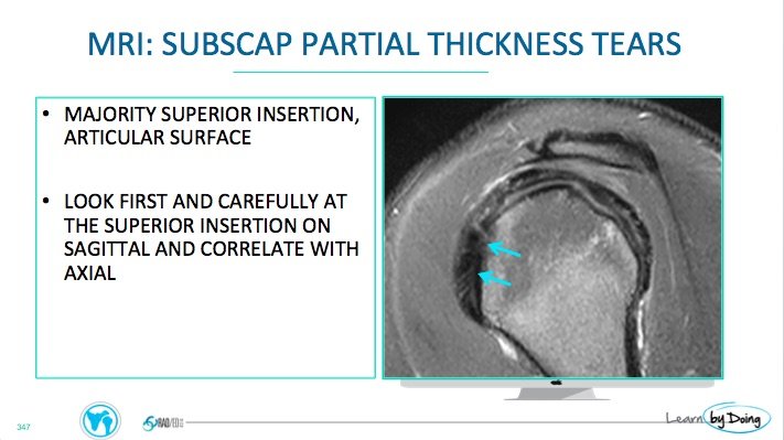
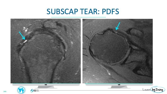 Image Above: Focal Partial tear at insertion ( Blue Arrow) articular surface. Note shape of humeral head which is flattened at the subscapularis insertion.
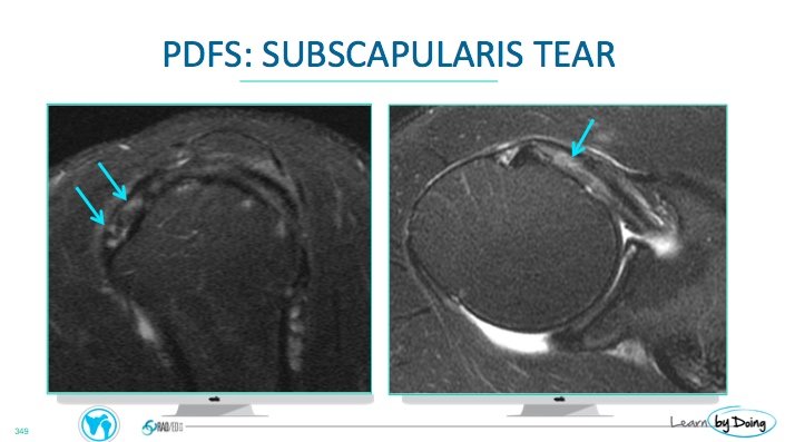
Image above: More extensive partial articular surface tears ( blue arrows) at expected location where subscapularis inserts on the flattened humeral head.
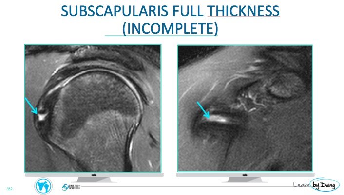
Image Above: Blue arrow full thickness tear ( incomplete, that is it only involves a part of the tendon)
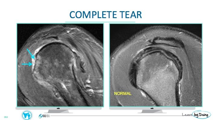
Image Above: Complete full thickness tear ( blue arrows). No fibres seen inserting onto lesser tuberosity ( bald man appearance).

