MRI Sacroileitis Chronic Changes
The chronic changes of sacroileitis on MRI. What are they and what do they look like.
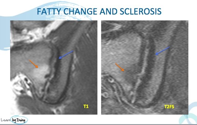
1 & 2 SUBCHONDRAL SCLEROSIS AND FATTY CHANGE
Image Above: Sclerosis at the margins of the SIJ ( blue arrow) and fatty deposits at sites of previous bone marrow oedema ( red arrow).
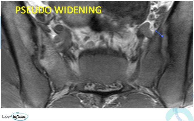
3. PSEUDO WIDENING
Image above: The joint looks enlarged due to erosions destroying bone and giving the pseudo widening appearance of the SI joint ( blue arrow).
4. BACK FILL
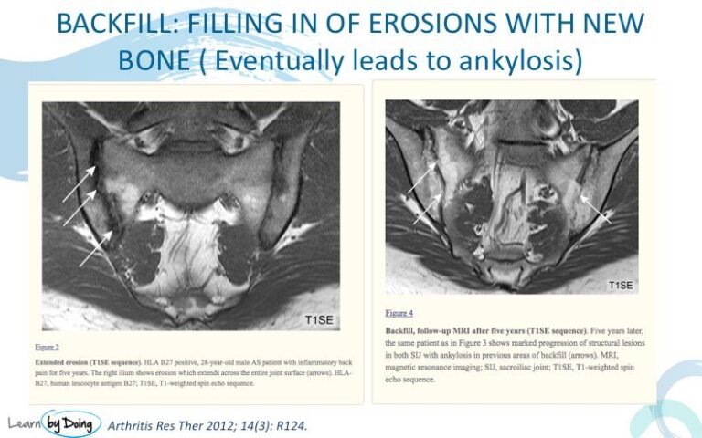 Image Above: White arrows in first image demonstrate erosions ( look particularly at the long sacral erosion. The second image is 5 years later where the sacral erosion on the right is now ” filled in” with bone ( fatty marrow signal) which si the backfill. This eventually leads to ankylosis across the joint.
5. ANKYLOSIS
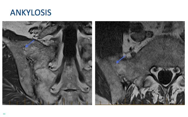 Image Above: The final stage of sacroileitis where there is fusion across the joint ( blue arrows).

