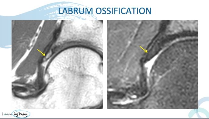
Hip Labrum Ossification MRI and Xray findings
Ossification of the acetabular labrum can be tricky to notice on MRI. The cases we have seen mostly have an underlying dysplastic, elongated labrum however there are reports of it occurring in a normal labrum.
So what does it look like? Having learnt the hard way by missing them by looking only at the MRI, I find the best place to start is with the plain xray as it can be overlooked if assessing just the MRI.
Image 2 Above: Bilateral labral ossification in the same patient. Labrum is diffusely ossified. Ossification follows the shape of the labrum. MRI below.
Image 3 Above: MRI for patient in Image 2. On the PD and PDFS scans ossification follows same signal as adjacent bone. Labrum is elongated and dysplastic.
Image 4 above: PD and PDFS ossification of the labrum centred on the labrum and follows labrum shape. Different patient to Image 2 and 3.


