Rotator Interval Capsulo Synovitis MRI
Rotator Interval Capsulitis Synovitis: In a previous post on this series of specific sites of synovitis in the shoulder, we looked at the Inferior Gleno Humeral Ligament. In this part, we look at the Rotator Interval of the shoulder. The anatomy of the Rotator Interval is quite complicated and requires another post to go through it properly, however, the three essential components that we assess for capsulitis/ synovitis, are the
- Coraco Humeral Ligament ( CHL) and the fat around the CHL
- Sub Coracoid fat
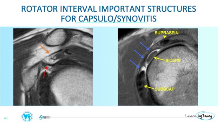
- Rotator Interval Capsule: This is the the anterior superior capsule between the Supraspinatus and Subscapularis tendons and merges with the CHL.
These are best assessed on sagittal PD non fat saturated scans.
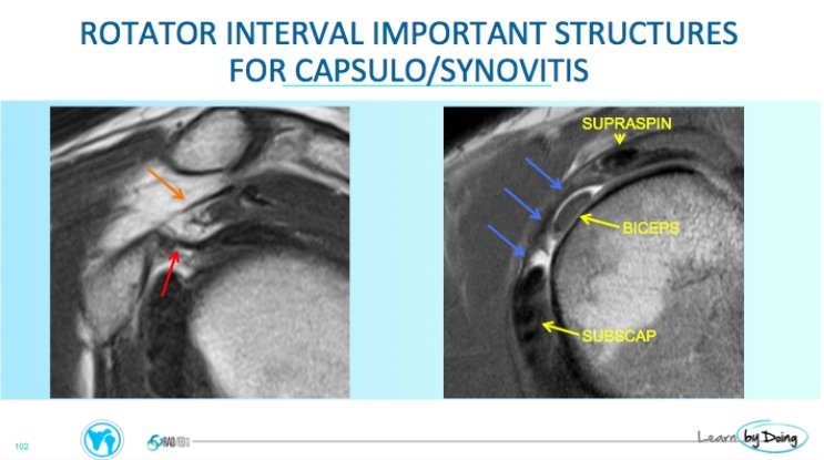 Image 1 above: Sagittal PD and PDFS: Red arrow- CoracoHumeral Ligament, Orange Arrow- Coraco Acromial Ligament, Blue Arrows- Rotator Interval Capsule
When the Rotator Interval is involved in Capsulo Synovitis, three things can occur
- Change in Fat signal: There is loss of the normal bright fat signal on PD around the CHL, replaced by lower signal inflammatory change. On the PDFS scans, instead of fat saturating and becoming dark, the fat is often bright. This can occur in the Subcoracoid Fat +/- the fat between the Coraco Acromial Ligament and the Coraco Humeral Ligament.
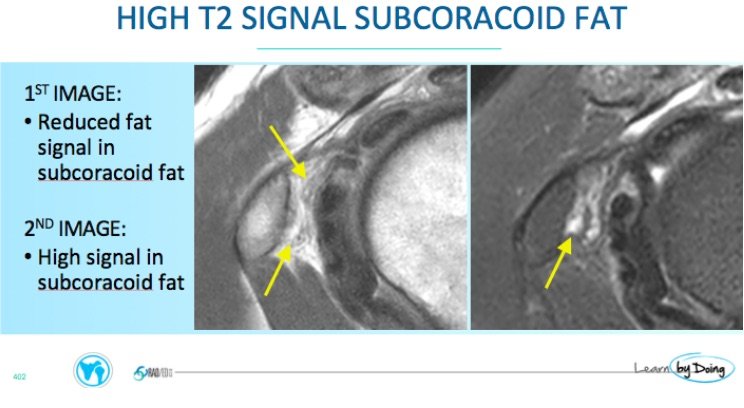 Image 2 above: PD and PDFS images: Increased signal on the PDFS image (right side yellow arrow) in the subcoracoid fat.
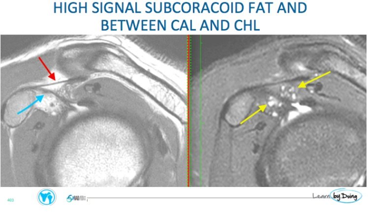 Image 3 above: Cystic type high T2 (Yellow Arrows) signal in Subcoracoid fat and in the fat between the Coraco Acromial ligament ( Red Arrow) and Coraco Humeral Ligament ( Blue Arrow) which is illdefined and slightly thick.
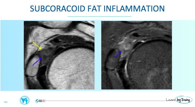 Image 4 above: Sagittal PD & PDFS : Diffuse inflammatory changes predominantly in subcoracoid fat ( Blue Arrow).CHL ( Yellow Arrow).
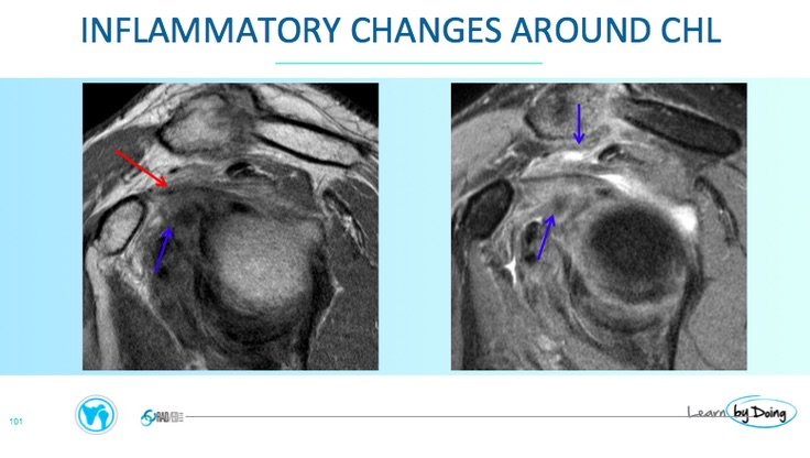 Image 5 above: Sagittal PD & PDFS : Diffuse inflammatory changes ( Blue arrow) above and below CHL ( Red Arrow) which is increased in intensity and ill defined ( Blue Arrow).
2. Thickening of the CHL ( Coraco Humeral Ligament): This is can be more subjective. In the workshops, we start with looking at multiple cases with normal CHLs and only then look at abnormal cases so that you have a reference point. So what does a normal CHL look like?
Normal CHL
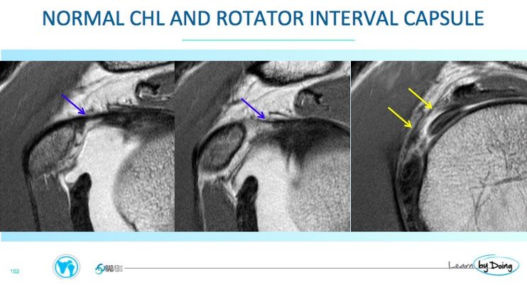 Image 6 above: Normal CHL ( Blue arrow) uniform low signal and sharp. Normal Anterior capsule at Rotator Interval ( Yellow Arrow).
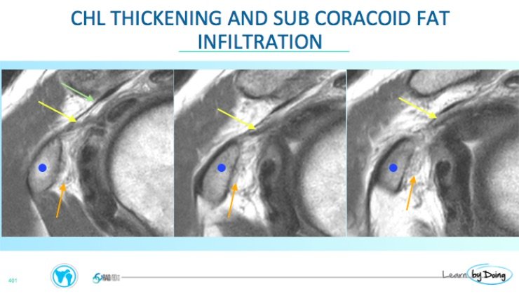 Image 7 above: Thickened and ill defined CHL ( Yellow arrow). Compare to normal Coraco Acromial Ligament ( Green Arrow) . Orange Arrow, infiltration of subcoracoid fat. Coracoid Process ( Blue Dot).
See also images 2, 3, 5 for examples of a thick CHL.
3. Thickening of the anterior capsule
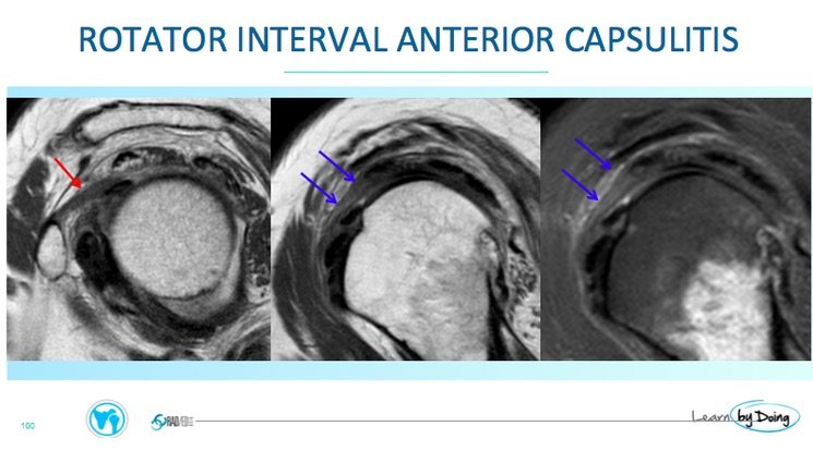 Image 8: Anterior capsular thickening ( Blue arrow). Compare with normal capsule in image 6. Thickening of the CHL ( Red arrow)


